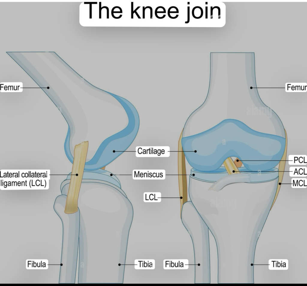
Have you ever wondered how your knee supports your entire body weight with such precision? The answer lies in the intricate femur tibia connection, a critical part of your knee anatomy. This connection is vital for movement and stability, making it one of the most fascinating aspects of human anatomy.
The femur, or thigh bone, acts as the primary connector between your hip and knee. It works in tandem with the tibia, the larger of the two bones in your lower leg, to form the knee joint. This joint is a synovial hinge, allowing for flexion and extension, as well as some rotational movements1.
Supporting this complex structure are essential components like the patella and fibula. The patella, or kneecap, slides along the femur’s surface, reducing friction and enhancing the strength of the knee joint1. Meanwhile, the fibula provides additional stability to the joint.
The knee joint itself is lubricated by synovial fluid, which reduces friction between the articulating surfaces of the femur and tibia. Articular cartilage covers these surfaces, ensuring smooth movement and shock absorption1. Proper alignment of these bones is crucial for joint function and overall body mechanics, directly influencing weight distribution and movement efficiency.
Understanding the femur tibia connection is the first step in appreciating the remarkable engineering of the human knee. This section will delve into the detailed anatomy of this vital joint, exploring how its various components work together to enable movement and maintain stability.
Key Takeaways
- The femur connects the hip to the knee, while the tibia links the knee to the ankle.
- The knee joint is a synovial hinge, allowing for flexion, extension, and limited rotation.
- The patella and fibula provide essential support to the knee joint.
- Synovial fluid and articular cartilage reduce friction and enable smooth movement.
- Proper bone alignment is crucial for joint function and overall body mechanics.
Introduction to the Femur Tibia Connection
The femur and tibia form a vital joint in the human body, essential for movement and stability. This connection is at the heart of the knee joint, enabling activities like walking, running, and climbing stairs. Understanding this joint is crucial for appreciating the intricacies of human anatomy.
Defining Key Structures
The femur, the longest and strongest bone in the body, connects the hip to the knee2. At the knee, it articulates with the tibia, the larger bone in the lower leg. The joint is a synovial hinge, allowing for flexion and extension, with some rotational movement possible when the knee is slightly flexed3.
The femur’s condyles—medial and lateral—sit atop the tibia’s plateaus, forming the joint. The medial condyle is larger and supports more body weight, while the lateral condyle prevents patellar dislocation3. Articular cartilage covers these surfaces, reducing friction and enabling smooth movement2.
The menisci, shock-absorbing structures within the knee, further enhance stability and reduce wear on the joint surfaces. Ligaments like the anterior and posterior cruciate ligaments add strength and stability to the joint3.
| Structure | Role | Contribution to Stability |
|---|---|---|
| Femur | Connects hip to knee | Provides structural support and leverage |
| Tibia | Supports body weight | Distributes weight and facilitates movement |
| Menisci | Shock absorption | Reduces joint wear and enhances stability |
| Ligaments | Joint stabilization | Prevents excessive movement and maintains joint integrity |
This intricate connection is vital for daily activities, making it a remarkable example of anatomical engineering.
Anatomy of the Knee Joint
The knee joint is a complex structure made up of four key bones: the femur, tibia, patella, and fibula. These bones work together to provide stability and enable movement. The femur, or thigh bone, is the longest and strongest bone in the body, while the tibia, or shinbone, is the weight-bearing bone of the lower leg4.
Bones Involved: Femur, Tibia, Patella, and Fibula
The femur connects to the tibia at the knee joint, forming a synovial hinge that allows for flexion, extension, and some rotation when slightly flexed4. The patella, or kneecap, slides along the femur’s surface, reducing friction and enhancing the strength of the knee joint4. The fibula, on the outside of the lower leg, provides additional stability to the joint.
The Role of Articular Cartilage and Menisci
Articular cartilage covers the ends of the femur and tibia, reducing friction and enabling smooth movement5. The menisci, located between the femur and tibia, act as shock absorbers and assist in rotating the knee. The menisci are composed of approximately 75% water by wet weight, with the outer one-third well-vascularized and the inner two-thirds avascular, affecting their healing potential5.
Tears in the menisci can cause pain, swelling, and limited mobility, often requiring medical attention6. Maintaining healthy joint surfaces is crucial for mobility and preventing conditions like osteoarthritis.
The Anatomy of the Femur
The femur, a marvel of anatomical engineering, serves as the cornerstone of the human lower limb, seamlessly integrating strength and flexibility to support both mobility and stability. As the longest and strongest bone in the body, it plays a pivotal role in weight-bearing and movement. Understanding its intricate structure is essential for appreciating its function in the human body.
Femoral Structure and Landmarks
The femur is divided into three main parts: the proximal (upper) end, the shaft, and the distal (lower) end. The proximal end features the femoral head, neck, and trochanters. The head forms a ball-and-socket joint with the hip, while the neck connects the head to the shaft7. The greater and lesser trochanters serve as attachment points for powerful muscles that facilitate hip movement.
The shaft, or diaphysis, is the long, cylindrical portion of the femur, designed for strength and durability. It is supported by a robust blood supply, primarily from the femoral artery, which branches into the medial and lateral circumflex arteries to nourish the femoral head7. The distal end includes the condyles, which articulate with the tibia to form the knee joint. The medial condyle is larger, bearing more body weight, while the lateral condyle helps prevent patellar dislocation8.
Muscle Attachments and Joint Function
Muscles attach to the femur via tendons and ligaments, enabling a wide range of movements. The quadriceps femoris muscle, innervated by the femoral nerve, attaches to the patella, which in turn connects to the tibia via the patellar tendon. This arrangement allows for powerful knee extension7. Conversely, the hamstring muscles, innervated by the sciatic nerve, facilitate knee flexion and hip extension8.
The femur also features the intertrochanteric line and crest, which are critical for muscle attachments. These landmarks provide anchorage points for muscles like the iliopsoas and gluteus minimus, essential for hip flexion and stabilization7. The femoral artery bifurcates near the lesser trochanter, supplying blood to the lower limb7.
| Structure | Role | Contribution to Stability |
|---|---|---|
| Femoral Head | Articulates with the hip joint | Enables hip movement and weight transfer |
| Condyles | Form the knee joint with the tibia | Supports body weight and facilitates movement |
| Trochanters | Muscle attachment points | Enhances movement through muscle leverage |
The femur’s structure is a testament to its vital role in human anatomy, providing the necessary support and mobility for daily activities. Its intricate landmarks and muscle attachments work in harmony to ensure efficient movement and stability.
The Importance of the Tibia in Lower Limb Stability
The tibia, often overshadowed by the femur, plays a crucial role in lower limb stability and weight distribution. As the second-largest bone in the body, it bears significant weight, making it indispensable for movement and balance9.
Weight Bearing and Shock Absorption
The tibia’s robust structure is designed to handle substantial weight. The medial aspect of the tibia supports the majority of the body’s weight, while the lateral aspect assists in balancing and shock absorption during activities like walking or running10.
Articular Surface and Meniscus Interaction
The tibial plateau, the upper surface of the tibia, forms the knee joint with the femur. Here, menisci act as shock absorbers, reducing friction and stabilizing the joint. This interaction is vital for smooth movement and preventing wear on the joint surfaces11.
| Structure | Role | Contribution to Stability |
|---|---|---|
| Tibial Plateau | Forms the knee joint with the femur | Supports body weight and facilitates movement |
| Menisci | Shock absorption and joint lubrication | Reduces wear and enhances stability |
| Ligaments | Joint stabilization | Prevents excessive movement and maintains integrity |
Proper alignment of the tibia is essential for joint function, preventing injuries and ensuring efficient movement. Understanding the tibia’s role is key to appreciating lower limb mechanics and overall mobility.
Ligaments and Tendon Attachments: Ensuring Knee Stability
The knee joint’s stability is largely attributed to its complex network of ligaments and tendons. These structures work in harmony to provide support and enable smooth movement. Understanding their roles is essential for appreciating the knee’s functionality.
Cruciate Ligaments (ACL & PCL) Dynamics
The anterior cruciate ligament (ACL) and posterior cruciate ligament (PCL) are vital for anterior and posterior stability, respectively. The ACL, often injured in sports, provides up to 85% of anterior stability when the knee is flexed12. PCL injuries commonly occur from direct impacts, such as in car accidents13.
Medial and Lateral Collateral Ligament Functions
The medial collateral ligament (MCL) and lateral collateral ligament (LCL) offer side stability. The MCL, more prone to injury, is often associated with medial meniscal tears due to its attachments8. The LCL, on the outer side, provides additional stability but is less frequently injured.
Ligament injuries can lead to pain, swelling, and instability. Treatment often starts with rest and ice, but severe cases may require surgery13. Recovery involves physical therapy to restore strength and mobility, ensuring the knee returns to optimal function.
For more detailed information on knee anatomy, visit this resource.
Exploring the Femur Tibia Connection
The femur and tibia work together to form one of the most essential joints in the human body, enabling movement and supporting body weight. This connection is crucial for activities like walking, running, and climbing stairs. Understanding this joint is vital for appreciating the intricacies of human anatomy.
Joint Mechanics Driving Movement
The femur, the longest and strongest bone in the body, connects the hip to the knee14. At the knee, it articulates with the tibia, the larger bone in the lower leg. The joint is a synovial hinge, allowing for flexion and extension, with some rotational movement possible when the knee is slightly flexed15.
The femur’s condyles—medial and lateral—sit atop the tibia’s plateaus, forming the joint. The medial condyle is larger and supports more body weight, while the lateral condyle prevents patellar dislocation15. Articular cartilage covers these surfaces, reducing friction and enabling smooth movement14.
The menisci, shock-absorbing structures within the knee, further enhance stability and reduce wear on the joint surfaces. Ligaments like the anterior and posterior cruciate ligaments add strength and stability to the joint15.

Understanding the femur tibia connection is the first step in appreciating the remarkable engineering of the human knee. This section will delve into the detailed anatomy of this vital joint, exploring how its various components work together to enable movement and maintain stability.
Key Takeaways
- The femur connects the hip to the knee, while the tibia links the knee to the ankle.
- The knee joint is a synovial hinge, allowing for flexion, extension, and limited rotation.
- The patella and fibula provide essential support to the knee joint.
- Synovial fluid and articular cartilage reduce friction and enable smooth movement.
- Proper bone alignment is crucial for joint function and overall body mechanics.
Importance of Proper Bone Alignment
Proper alignment of the femur and tibia is essential for joint function and overall body mechanics. Misalignment can lead to increased wear on the articular surfaces, potentially causing pain and dysfunction. For example, fractures of the femur or tibia can significantly impact knee function and mobility16.
Research findings highlight the connection between alignment and knee performance. Optimal alignment is critical for injury prevention and maintaining efficient movement14.
Clinical Implications and Common Knee Disorders
Knee disorders are a common issue affecting millions worldwide, leading to pain, limited mobility, and reduced quality of life. Understanding these conditions is crucial for early diagnosis and effective treatment.
Fractures, Ligament Tears, and Joint Injuries
Fractures in the femur or tibia can significantly impact mobility and often require surgical intervention17. Ligament tears, particularly in the ACL, are prevalent in sports, with women being four to eight times more likely to suffer ACL injuries than men18. These injuries often present with a popping sound, followed by swelling and instability17.
Degenerative Conditions and Arthritis Overview
Osteoarthritis is a leading cause of knee pain, especially in older adults, and is exacerbated by obesity and aging18. It causes cartilage degradation, leading to stiffness and loss of function. Conservative treatments like weight reduction and physical therapy are often recommended before considering surgery18.
Early diagnosis is key to managing these conditions effectively. Imaging tools such as MRIs and X-rays are commonly used for accurate assessments17. Prompt treatment can prevent further complications and restore knee function.
Surgical Interventions and Rehabilitation Strategies
When conservative treatments fail, surgical interventions become necessary to restore joint function and alleviate pain. Modern orthopedic procedures have evolved significantly, offering effective solutions for various knee-related injuries and conditions. Rehabilitation plays a crucial role in post-surgical recovery, ensuring patients regain strength and mobility.
| Procedure | Description | Success Rate |
|---|---|---|
| ACL Reconstruction | Reconstructing the anterior cruciate ligament, often using patellar tendon grafts19. | 85-90% success rate in restoring stability20. |
| Total Knee Replacement | Replacing the damaged joint with artificial components. | 90-95% success rate in pain relief and improved function19. |
| Minimally Invasive Surgery | Less invasive techniques to repair fractures or ligaments. | High success rate with faster recovery times20. |
Post-surgical recovery involves physical therapy to restore joint mobility and strength. Studies show that patients who undergo rigorous physical therapy achieve better outcomes, with significant improvements in knee flexion and extension20. A multidisciplinary rehabilitation team, including physical therapists and orthopedic specialists, is essential for optimal recovery.
The Role of Biomechanics in Joint Function
Biomechanics plays a vital role in understanding how joints function under various loads and movements. The study of biomechanics helps us appreciate how forces are distributed across the knee joint, ensuring efficient movement and minimizing the risk of injury. This section explores how biomechanical principles influence joint loading and efficiency, drawing on research findings regarding weight distribution and movement mechanics.
Understanding Weight Distribution and Movement Efficiency
The distribution of weight across the knee joint is a critical aspect of biomechanics. During activities like walking or running, the medial femoral condyle supports the majority of the body’s weight, while the lateral condyle assists in balancing and shock absorption. Research indicates that the medial femoral condyle translates no more than 7.1 mm anteroposteriorly during weight-bearing activities21, while the lateral femoral condyle translates posteriorly about 15 mm during flexion21.
Bone geometry and alignment significantly influence movement efficiency. The articular surfaces of the femur and tibia are designed to reduce friction and distribute forces evenly. For instance, the average radius of the medial femoral condyle’s articular surface is about 22 mm, subtending an arc of 110°21. This design ensures smooth movement and reduces wear on the joint surfaces.
The relationship between force, load-bearing, and joint health is well-documented. Proper biomechanics contribute to long-term joint stability and reduce the risk of injuries. Studies show that biomechanical efficiency correlates with reduced injury risk, as it ensures forces are distributed optimally across the joint22.
In conclusion, understanding biomechanics is essential for improving clinical outcomes and maintaining healthy joints. By studying how forces interact with joint structures, we can develop better treatments and rehabilitation strategies to enhance movement efficiency and reduce injury risk.
Conclusion
Understanding the femur tibia connection is crucial for appreciating how the knee supports body weight and enables movement. This joint is essential for activities like walking and running, making it a cornerstone of human anatomy.
The femur and tibia work together to form a synovial hinge joint, allowing for flexion and extension. The femur’s condyles sit atop the tibia’s plateaus, with articular cartilage reducing friction and enabling smooth movement23. The menisci act as shock absorbers, further enhancing stability.
Proper bone alignment is vital for joint function. Misalignment can lead to increased wear on joint surfaces, potentially causing pain and dysfunction. For instance, fractures of the femur or tibia can significantly impact knee function and mobility24.
Knee disorders, such as ligament tears and osteoarthritis, are common issues affecting millions. Early diagnosis is key to effective treatment, which may include surgical interventions like ACL reconstruction or total knee replacement25.
At our organization, we are committed to advancing the understanding and treatment of orthopedic conditions. We encourage readers to explore further resources and consult with specialists for more in-depth information. Maintaining joint health is essential for a better quality of life.
