When it comes to diagnosing a medial collateral ligament (MCL) injury, accuracy is key. The MCL, a crucial ligament in the knee, plays a vital role in stability. Injuries to this area can significantly impact mobility and overall knee function.
Doctors employ a combination of physical examinations and imaging tests to assess the severity of an MCL injury. From hands-on evaluations to advanced imaging techniques, these methods ensure a precise diagnosis, which is essential for effective treatment and recovery.
In this article, we will guide you through the diagnostic procedures used to identify MCL injuries. We will explore the role of doctors in performing these tests and the importance of accurate diagnosis for proper treatment.
Key Takeaways
- Understanding the MCL and its role in knee stability.
- Physical examination techniques used by doctors.
- Imaging tests for accurate diagnosis.
- Importance of early and precise diagnosis.
- Realistic images and clear guidance for better understanding.
Understanding Medial Collateral Ligament Injuries
The medial collateral ligament (MCL) is a critical structure in the knee, providing stability and support during various activities. As a key ligament, it plays a vital role in connecting the femur (thigh bone) to the tibia (shin bone), ensuring proper knee function and movement.
What is the MCL and Why is it Important?
The MCL is one of the four major ligaments in the knee, specifically located on the medial (inner) side. It works in tandem with the anterior cruciate ligament (ACL) and other knee structures to maintain stability, especially during sideways movements and twisting actions.
Common Causes of MCL Injury in Sports and Daily Life
MCL injuries often occur due to direct blows to the outer knee, sudden twists, or repetitive stress from high-impact activities. Sports like football, soccer, and basketball, where sudden changes in direction are common, pose a higher risk. Even everyday activities, such as slipping on uneven surfaces, can lead to MCL tears.
| Aspect | Details |
|---|---|
| MCL Function | Provides medial knee stability |
| Common Causes | Sports collisions, twisting motions |
| Risk Factors | High-impact sports, repetitive stress |
Understanding the MCL’s role and the common causes of injury is essential for prevention and effective treatment. By recognizing the risks, individuals can take proactive steps to protect their knees and maintain optimal joint health.
How Do Doctors Test for MCL Injury
A thorough physical examination is the first step in assessing a suspected MCL tear. During this process, healthcare professionals evaluate the knee’s stability and look for signs of injury.
Physical Examination: Hands-on Testing Techniques
Doctors use several hands-on methods to diagnose MCL injuries. These include palpation to identify areas of tenderness and stress tests to assess ligament stability. A common test is the valgus stress test, where the knee is gently bent outward to check for excessive movement or pain.
Symptoms such as pain, swelling, or a “popping” sensation are carefully noted. These observations help determine the severity of the injury, ranging from mild sprains to complete tears. The findings guide further diagnostic steps and treatment plans.
| Test Type | Description |
|---|---|
| Palpation | Identifies areas of tenderness and swelling. |
| Valgus Stress Test | Assesses medial ligament stability by bending the knee outward. |
| Varus Stress Test | Evaluates lateral ligament stability by bending the knee inward. |
These hands-on techniques provide valuable insights into the extent of the injury, helping to differentiate between mild and severe cases. Accurate diagnosis is crucial for effective treatment and recovery.
Diagnostic Imaging: Enhancing Accuracy
Accurate diagnosis of medial collateral ligament injuries often requires more than a physical exam. Diagnostic imaging plays a crucial role in confirming the extent of the damage and guiding treatment plans.
X-Ray and Stress X-Ray Procedures
X-rays are typically the first imaging tests used. They help identify bone fractures or misalignments in the knee joint. Stress X-rays, on the other hand, involve bending the knee under stress to assess ligament stability. These tests are particularly useful for detecting joint instability that may not be visible on a standard X-ray.
Magnetic Resonance Imaging (MRI) Insights
MRI provides detailed images of soft tissues, including ligaments, tendons, and cartilage. It is especially effective in diagnosing complete or partial MCL tears. MRI can also reveal swelling inside the knee and assess the severity of any bone or cartilage damage.
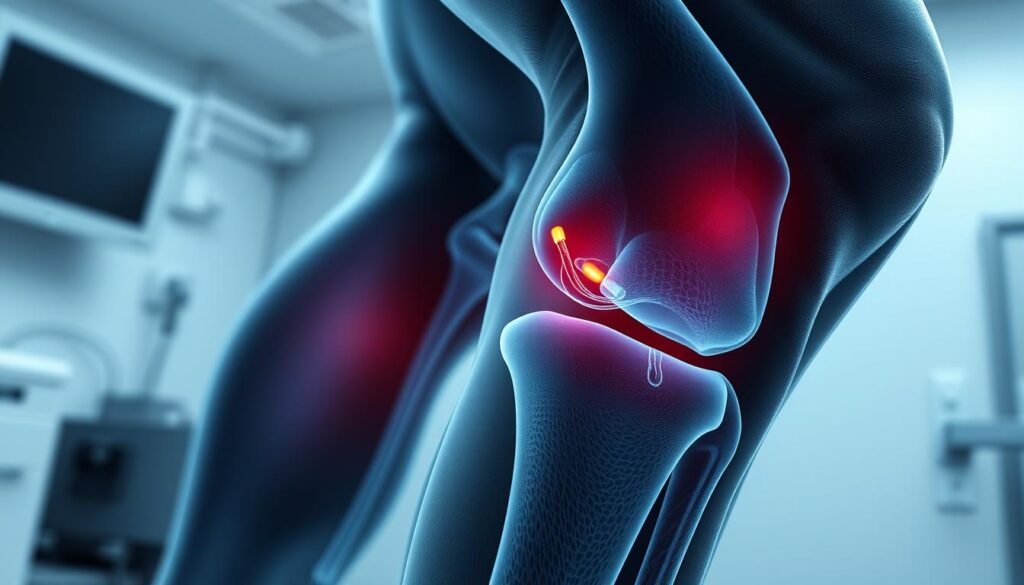
Realistic imaging techniques enhance diagnostic accuracy by providing clear visuals of the knee’s internal structures. This helps in identifying signs like swelling, which can confirm a diagnosis. By combining these imaging methods, healthcare professionals can develop a comprehensive treatment plan tailored to the patient’s needs.
Assessing Injury Severity and Grading
Understanding the severity of a medial collateral ligament tear is crucial for effective treatment. The injury is graded based on the extent of ligament damage, which directly impacts recovery strategies and outcomes.
Understanding Grade 1, Grade 2, and Grade 3 Tears
A Grade 1 tear involves mild stretching of the ligament, causing minimal pain and no instability. Recovery typically occurs within a few weeks with conservative treatment.
A Grade 2 tear signifies a partial ligament tear, leading to moderate pain and some joint laxity. Recovery may take several weeks, often requiring bracing and physical therapy.
A Grade 3 tear is the most severe, involving a complete ligament tear. This results in significant instability and pain, often requiring surgical intervention and an extended recovery period.
Accurate grading is essential to determine the best course of action, ensuring proper healing and preventing further complications. Each grade presents unique challenges, making a thorough assessment vital for effective treatment planning.
Non-Surgical Testing Methods and Treatment Options
When addressing medial collateral ligament injuries, non-surgical approaches are often the first line of defense. These methods focus on reducing pain, promoting healing, and restoring knee function without the need for invasive procedures.
Examining the RICE Protocol and Pain Management
The RICE protocol—Rest, Ice, Compression, and Elevation—is a cornerstone of initial treatment. Rest helps avoid further strain, while ice reduces swelling. Compression and elevation further minimize inflammation. For pain management, nonsteroidal anti-inflammatory drugs (NSAIDs) like ibuprofen are commonly prescribed to alleviate discomfort and inflammation.
Role of Physical Therapy and Knee Bracing
Physical therapy plays a crucial role in rehabilitation. Tailored exercises improve strength, flexibility, and range of motion. Knee bracing provides additional support, stabilizing the joint during recovery. Crutches may also be recommended to reduce stress on the knee, allowing the ligament to heal properly without additional strain.
Combining these non-surgical strategies creates a comprehensive treatment plan, often avoiding the need for surgery. Early intervention and adherence to these methods can significantly enhance recovery outcomes.
Surgical Considerations for Severe Cases
In severe cases where non-surgical methods are insufficient, surgery becomes the necessary intervention for MCL tears. This is particularly true when the injury is combined with other ligament damage, such as an ACL tear, leading to significant knee instability.
When and Why Surgery May Be Recommended
Surgery is typically recommended for Grade 3 MCL tears, where the ligament is completely torn, or when there is concurrent damage to other knee structures like the ACL. The procedure often involves reconstructing the ligament using grafts, which can be taken from the patient’s body or from a donor.
The surgical process typically involves arthroscopy or open surgery, depending on the injury’s complexity. Post-operative care is crucial, with realistic images and illustrations guiding patients through recovery steps to ensure proper healing and prevent complications.
Accurate diagnosis plays a vital role in determining the need for surgery. By combining advanced imaging techniques with thorough physical examinations, healthcare professionals can identify cases that require surgical intervention, ensuring the best possible outcomes for patients.
Rehabilitation and Recovery Strategies
Recovering from an MCL injury requires a well-structured approach to ensure proper healing and restore knee function. Our rehabilitation programs are tailored to address each patient’s specific needs, focusing on strength, flexibility, and joint stability.
Customized Physical Therapy Approaches
Physical therapy is a cornerstone of MCL recovery. Therapists design personalized exercise plans to enhance strength and improve knee mobility. Early stages focus on restoring range of motion, while later phases incorporate resistance training and balance exercises.
Each patient works closely with a therapist to progress through exercises safely, avoiding re-injury. This collaborative approach ensures a comprehensive recovery plan.
Expected Recovery Timelines and Home Care Tips
Recovery time varies based on injury severity. Grade 1 injuries may heal within 2-4 weeks, while Grade 3 tears could take 3-6 months. Consistency in therapy and adherence to home care routines are crucial.
At home, patients should continue prescribed exercises, use braces for support, and monitor their progress. Realistic expectations and patience are essential for full recovery.
| Grade | Weeks | Knee Function | Activities |
|---|---|---|---|
| 1 | 2-4 | Mild limitation | Light exercises |
| 2 | 6-8 | Moderate limitation | Strengthening exercises |
| 3 | 12-24 | Significant limitation | Advanced rehab |
Realistic Imaging Techniques and Their Benefits
Imaging plays a vital role in accurately diagnosing MCL injuries, and advancements in this field have revolutionized how we assess knee injuries. Realistic imaging provides clear visuals of the knee’s internal structures, aiding precise diagnosis and treatment planning.
Advancements in Diagnostic Imaging
Recent advancements in imaging techniques have significantly improved the detection of swelling and subtle joint changes. High-resolution MRI and CT scans offer detailed views of the MCL, allowing doctors to identify even minor tears. These images are captured from multiple angles, including the side and inside of the knee, to provide a comprehensive view of the injury.
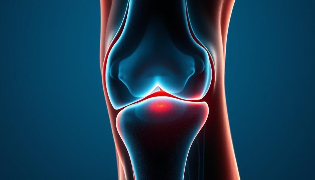
The ability to view the knee from different angles, such as the side, helps in pinpointing the exact location of the tear. Inside views, on the other hand, reveal swelling and ligament damage that might be missed in standard images. This detailed visualization is crucial for developing effective treatment plans.
Realistic imaging also helps in monitoring the healing process. By comparing images over time, healthcare providers can track the reduction in swelling and the recovery of the MCL. This approach ensures that the treatment plan is adjusted as needed for optimal results.
In conclusion, realistic imaging techniques have become essential in diagnosing and managing MCL injuries. Their ability to provide detailed, high-resolution images from various angles, including the side and inside of the knee, ensures accurate diagnoses and effective treatment plans.
Our Approach: Integrating Testing with Personalized Care
At our center, we pride ourselves on a comprehensive approach that combines advanced diagnostic techniques with tailored treatment plans. Our team of specialists works collaboratively to ensure each patient receives care that addresses their unique needs.
Team-Based Strategies and Tailored Treatment Plans
Our doctors employ a combination of physical examinations and cutting-edge imaging to ensure accurate diagnoses. From stress tests to MRI scans, every tool is used to gather detailed insights into the injury’s severity. This thorough approach allows us to craft personalized treatment plans that target the specific aspects of each MCL tear or injury.
How We Ensure Accurate Diagnosis and Effective Recovery
Accuracy is paramount in our practice. By integrating hands-on exams with advanced imaging, we ensure that every diagnosis is precise. Our treatment plans are designed to promote healing, restore function, and prevent future injuries. Whether it’s through physical therapy, bracing, or surgery, we tailor our strategies to meet each patient’s specific recovery needs.
Our collaborative approach involves a team of specialists, ensuring comprehensive care. From initial testing to rehabilitation, we guide patients through every step of their journey, fostering trust and confidence in their recovery process.
Tips for Preventing Future MCL Injuries
Preventing future MCL injuries requires a proactive approach that combines targeted exercises, proper warm-up routines, and mindful activity modification. By incorporating these strategies into your daily routine, you can significantly reduce the risk of experiencing another injury.
Strengthening Exercises and Proper Warm-Up Routines
Strengthening the muscles around the knee, particularly the thigh, is essential for preventing MCL injuries. Exercises like squats, lunges, and leg presses can help build strength and stability. Additionally, incorporating balance and flexibility exercises into your routine can improve overall knee function and reduce injury risk.
A proper warm-up before any sport or physical activity is crucial. It prepares the muscles and joints for the upcoming demands, reducing the likelihood of strains or tears. A dynamic warm-up that includes movements like high knees, leg swings, and calf raises can effectively prepare the body for activity.
| Exercise Type | Frequency | Benefits |
|---|---|---|
| Squats | 3 times a week | Strengthens thigh muscles and improves knee stability |
| Lunges | 3 times a week | Enhances balance and overall lower body strength |
| Leg Press | 2 times a week | Targets multiple muscle groups for comprehensive strength |
Incorporating these exercises into your weekly routine can make a significant difference in injury prevention. For more detailed information on MCL injuries and prevention strategies, visit Cleveland Clinic’s resource on MCL tears.
Conclusion
In conclusion, addressing medial collateral ligament tears requires a blend of accurate diagnosis, personalized treatment, and proactive care. Understanding the severity of the tear is vital for determining the best course of action, whether through non-invasive methods or surgical intervention. Our team emphasizes a tailored approach, combining realistic imaging with customized therapy to ensure optimal recovery.
Realistic imaging techniques have revolutionized how we diagnose and manage MCL tears. By providing detailed visuals from multiple angles, these methods enhance diagnostic accuracy and guide effective treatment plans. Our commitment to personalized care ensures that each patient receives a treatment plan that addresses their unique needs, promoting faster healing and restoring knee function.
If you’re experiencing symptoms like knee pain or instability, seek prompt medical evaluation to prevent further complications. Early diagnosis and appropriate treatment can significantly improve outcomes. For ongoing knee health, incorporate strengthening exercises, proper warm-ups, and mindful activity modifications into your routine.
Take the first step towards recovery by consulting with our specialists. With a focus on comprehensive care and advanced diagnostic techniques, we are here to guide you through every phase of your journey. Remember, proactive care and personalized treatment are key to maintaining healthy knees and preventing future issues.
FAQ
What are the common symptoms of an MCL tear?
Common symptoms include pain on the inner side of the knee, swelling, and difficulty bending or straightening the knee. Some people may also experience tenderness or instability in the joint.
How long does it take to recover from an MCL injury?
Recovery time varies based on the grade of the tear. Grade 1 injuries may heal in a few weeks, while Grade 3 tears could take several months. Proper physical therapy and rest are key to a full recovery.
Can an MCL tear heal without surgery?
Yes, most MCL tears heal without surgery. Physical therapy, bracing, and the RICE protocol (rest, ice, compression, elevation) are often effective for non-severe cases. Surgery is typically reserved for severe or complex injuries.
What role does physical therapy play in MCL recovery?
Physical therapy is crucial for restoring strength, flexibility, and stability to the knee. A personalized exercise program can help prevent future injuries and improve overall joint function.
How can I prevent an MCL injury?
Strengthening the thigh and leg muscles, warming up properly before sports, and using proper techniques during activities can help reduce the risk of an MCL injury.
What imaging tests are used to diagnose an MCL tear?
MRI is the most accurate imaging test for diagnosing an MCL tear. X-rays may also be used to rule out bone fractures or other joint issues.
Do I need a knee brace after an MCL injury?
A knee brace may be recommended to provide stability and support during the healing process, especially in more severe cases or during physical activity.
Can I return to sports after an MCL injury?
Yes, but it’s important to wait until the knee has fully healed and you’ve regained strength and stability. Your healthcare provider will help determine when it’s safe to return to sports.
What are the grades of MCL tears?
MCL tears are classified into three grades: Grade 1 (mild stretch), Grade 2 (partial tear), and Grade 3 (complete tear). The treatment approach varies based on the grade.
How does an MCL injury differ from an ACL injury?
The MCL and ACL are both ligaments in the knee, but they are located in different areas. The MCL is on the inner side, while the ACL is in the center. Symptoms and treatment may overlap but are not identical.
Can I use crutches after an MCL injury?
In some cases, crutches may be recommended to avoid putting weight on the injured knee, especially during the initial healing phase. Your doctor will advise based on the severity of the injury.
Pingback: How to test for MCL sprain at home?