Have you ever wondered if an MCL tear is painful to touch? This is a common question for many individuals dealing with knee injuries. The medial collateral ligament (MCL) is a crucial ligament on the inner side of the knee, and injuries to this area can significantly impact daily activities.
When an MCL tear occurs, it can range from a mild sprain to a complete tear. One of the primary concerns for those experiencing this injury is the level of pain involved. While some may feel a sharp, immediate pain, others might notice discomfort that develops over time.
Common symptoms of an MCL tear include a popping sensation at the time of injury, swelling, and bruising around the inner knee. These symptoms can make it challenging to bear weight or move the knee normally. Understanding these signs is crucial for proper diagnosis and treatment.
In this article, we will explore the symptoms of an MCL tear, diagnostic methods, treatment options (both surgical and non-surgical), and recovery strategies. We will also discuss how realistic images can help enhance your understanding of this injury.
Key Takeaways
- An MCL tear can cause pain and impair weight-bearing abilities.
- Symptoms include a popping sensation, swelling, and bruising.
- Treatment options range from non-surgical methods to surgery, depending on the severity.
- Proper diagnosis is essential for effective treatment.
- Visual aids can significantly enhance understanding of the injury and recovery process.
Understanding the Medial Collateral Ligament and MCL Tears
The medial collateral ligament (MCL) is a vital structure in the knee, providing essential stability. Located on the inner side of the knee, it connects the femur (thigh bone) to the tibia (shin bone), preventing excessive lateral movement. This ligament is crucial for maintaining proper knee alignment and supporting the joint during activities like walking or running.
The MCL is distinct from other knee ligaments due to its location and function. It is one of the four major ligaments in the knee, alongside the anterior cruciate ligament (ACL), posterior cruciate ligament (PCL), and lateral collateral ligament (LCL). The MCL’s unique position allows it to absorb shock and stress, making it prone to injuries during direct blows to the knee or sudden twists.
The MCL’s robust blood supply is a key factor in its healing process. Unlike some other ligaments, the MCL benefits from a rich vascular network, which promotes recovery and reduces the need for surgical intervention in many cases. This natural healing capacity is why many MCL tears can be effectively managed with non-surgical treatments, such as rest, ice, and physical therapy.
MCL tears can vary significantly in severity, ranging from minor sprains to complete ruptures. Mild tears may cause minimal discomfort and allow for continued activity, while severe tears can lead to instability and require more intensive treatment. Understanding the extent of the injury is crucial for developing an appropriate treatment plan.
Realistic images and anatomical diagrams play a significant role in helping individuals comprehend the nature of MCL tears. Visual aids can illustrate the ligament’s position, the severity of the tear, and the healing process, making complex medical information more accessible and easier to understand.
Recognizing Symptoms of an MCL Tear
Identifying the symptoms of an MCL tear is crucial for early diagnosis and effective treatment. The medial collateral ligament (MCL) is located on the inner side of the knee, and injuries to this area can present with distinct signs that help in understanding the severity of the condition.
Pain at the Inner Knee
Pain is often the most noticeable symptom of an MCL tear. This discomfort is usually localized to the inner knee area and can vary in intensity. Some individuals may experience a sharp, immediate pain at the moment of injury, while others might notice a gradual increase in discomfort. The pain can worsen with activities that involve twisting or bending the knee.
Swelling and Bruising Indicators
Swelling and bruising around the inner knee are common indicators of an MCL tear. These symptoms can develop soon after the injury and may persist for several days. The swelling can make the knee appear larger than usual, and the bruising may spread to the surrounding areas, depending on the severity of the tear.
The “Popping” Sensation
A significant indicator of an MCL tear is a “popping” sensation at the time of injury. This sound is often accompanied by immediate pain and can be a clear sign that something is wrong with the ligament. It’s important to seek medical attention if this occurs, as it may indicate a more severe injury.
Differentiating Pain Levels
It’s essential to differentiate between mild discomfort and more serious injury-related pain. Mild MCL tears might allow for continued activity with some pain, while more severe tears can lead to instability and difficulty in moving the knee. Understanding these differences can help in determining the appropriate course of action.
Impact on Knee Function
An MCL tear can significantly affect knee function, making it challenging to bear weight or move the knee normally. This impairment can interfere with daily activities and athletic performance, emphasizing the need for proper diagnosis and treatment.
Realistic visual representations, such as images and diagrams, can enhance understanding of the injury and its symptoms. These visuals provide a clearer picture of the ligament’s condition and the healing process, making complex medical information more accessible.
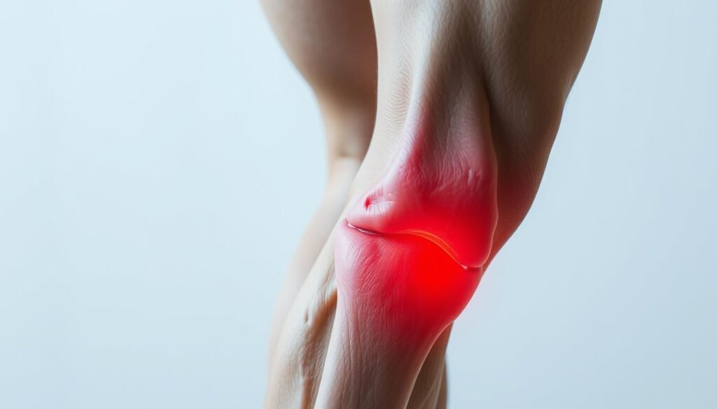
is mcl tear painful to touch? Our Insight
Understanding whether an MCL tear is painful to touch is crucial for those dealing with knee injuries. The medial collateral ligament, located on the inner side of the knee, plays a vital role in stability. When injured, it can cause significant discomfort.
Why does touching an MCL tear hurt so much? The ligament is richly supplied with nerves, making it sensitive. Even minor tears can cause discomfort when touched, and more severe injuries may lead to instability and pain during movement.
While some people might experience sharp pain immediately, others may notice discomfort that develops over time. It’s important to seek medical attention if pain persists, as it could indicate a need for further treatment.
MRI imaging is a valuable tool for confirming the extent of an MCL tear. It provides detailed images that help guide treatment decisions, ensuring the best possible outcome for recovery.
Assessing the Grades of MCL Injuries
Understanding the severity of an MCL injury is crucial for effective treatment. Medical professionals classify MCL tears into three grades based on the extent of the ligament damage.
Grade 1 & Grade 2 MCL Tears
A Grade 1 MCL tear involves minimal damage to the ligament fibers, causing slight pain and stiffness. It often allows for continued activity with minimal discomfort. A Grade 2 tear, however, involves partial ligament disruption, leading to moderate instability and noticeable pain during movement.
Considerations for a Grade 3 Tear
A Grade 3 MCL tear is the most severe, involving a complete rupture of the ligament. This results in significant instability, making it difficult to bear weight or move the knee. Grade 3 tears may also be accompanied by other structural damage, requiring more intensive treatment.
| Grade | Description | Symptoms |
|---|---|---|
| Grade 1 | Mild sprain with minimal fiber damage | Slight pain, minimal stiffness |
| Grade 2 | Partial tear with moderate instability | Noticeable pain, limited movement |
| Grade 3 | Complete tear with significant instability | Severe pain, inability to bear weight |
Causes and Risk Factors for MCL Tears
MCL tears often result from a combination of traumatic incidents and repetitive stress. Understanding these causes can help in preventing such injuries and managing their impact.
Sports and High-Impact Activities
High-impact sports like football and soccer significantly increase the risk of MCL tears. Sudden stops, quick turns, and twisting motions can strain the ligament, leading to injury. These sports often involve direct blows to the knee, which can cause immediate damage.
Overuse and Repetitive Stress
Repetitive stress from activities like running or cycling can gradually weaken the MCL. Over time, this stress may lead to a tear, especially if proper rest and recovery are not prioritized.
Risk Factors
Improper warm-ups and inadequate knee support are key risk factors. Both acute trauma and chronic overuse can lead to MCL tears, highlighting the importance of preventive measures.
Addressing these causes through awareness and proper techniques can reduce the likelihood of MCL tears and promote overall knee health.
How MCL Tears are Diagnosed
Diagnosing an MCL tear involves a combination of physical examinations and advanced imaging techniques. Doctors use these methods to assess the severity of the injury and determine the appropriate treatment plan.
Physical Examination Techniques
During a physical exam, a doctor will typically perform stress tests to evaluate knee stability. These tests involve bending or straightening the knee to check for ligament laxity. The doctor will also assess the tibia and femur for any abnormalities, as these bones are connected by the MCL. The examination may reveal instability or tenderness, which are key indicators of a tear.
Imaging Methods (MRI, X-ray, Ultrasound)
Imaging tests are crucial for confirming the diagnosis and assessing the extent of the tear. An MRI is the most detailed imaging method, providing clear visuals of the ligament and surrounding tissues. X-rays are often used to rule out fractures or bone misalignment, while ultrasounds offer a real-time view of the joint and ligaments. These imaging methods help doctors identify subtle findings that indicate the level of damage to the MCL.
Modern imaging advances have significantly improved the accuracy of MCL tear diagnoses. By evaluating the tibia, femur, and joint stability, doctors can develop a comprehensive treatment plan tailored to the patient’s needs.
Non-Surgical Treatments for MCL Tears
Non-surgical treatments are often the first line of defense for managing MCL tears, especially in less severe cases. These methods focus on reducing pain, swelling, and promoting natural healing.
RICE Method and Pain Management
The RICE method—Rest, Ice, Compression, and Elevation—is a cornerstone of non-surgical treatment. Rest helps avoid further injury, while ice reduces swelling. Compression with an elastic bandage and elevation above heart level also aid in minimizing swelling. For pain relief, nonsteroidal anti-inflammatory drugs (NSAIDs) like ibuprofen are commonly recommended.
Knee Bracing and Crutch Support
Knee braces provide stability and protection during the healing process, while crutches can help avoid putting weight on the injured knee. These supportive devices are especially useful in the early stages of recovery, allowing the ligament to heal without additional strain.
| Treatment Option | Details |
|---|---|
| RICE Method | Rest, Ice, Compression, Elevation to reduce pain and swelling. |
| NSAIDs | Pain management to alleviate discomfort. |
| Knee Brace | Provides stability and support during healing. |
| Crutches | Assist in mobility without putting weight on the knee. |
Non-surgical treatments are effective due to the MCL’s good blood supply, which promotes healing. Recovery for minor tears typically takes a few weeks, but it’s crucial to follow a doctor’s advice to ensure proper healing and prevent further issues. Realistic images can also help patients better understand their injury and treatment process.
Surgical Treatment Options for Severe MCL Injuries
In severe cases, surgery is often necessary to address MCL injuries. This intervention is typically reserved for Grade 3 tears or when the injury is accompanied by damage to other ligaments or bones.
Doctors may recommend surgery in cases where the ligament is completely ruptured or when non-surgical treatments fail to provide sufficient healing. The primary goal of surgery is to restore knee stability and prevent further complications.
| Procedure | Description |
|---|---|
| Ligament Reattachment | Reattaching the torn ligament to the bone using sutures or anchors. |
| Ligament Reconstruction | Replacing the damaged ligament with a graft, often taken from another part of the body or a donor. |
The surgical process typically involves small incisions to minimize tissue damage. Surgeons may use grafts to strengthen the repair. Recovery after surgery includes several weeks of immobilization, followed by physical therapy to restore knee function.

While most MCL injuries heal without surgery, severe cases require surgical intervention to ensure proper healing and prevent long-term instability.
The Importance of Physical Therapy in MCL Recovery
Physical therapy plays a vital role in restoring strength and mobility after an MCL injury. It helps patients regain normal joint function and reduces the risk of future injuries.
Rehabilitation Exercises
Targeted exercises are designed to strengthen the muscles around the knee, including those near the femur and thigh bone. These exercises improve joint stability and range of motion. For example, straight leg raises and hamstring stretches are often recommended.
Restoring Movement and Stability
Physical therapy focuses on restoring proper motion to the knee joint. This helps in speeding up the recovery process. A therapist may use exercises like step-ups or balance training to enhance stability and flexibility.
Professional therapists create personalized exercise programs tailored to each patient’s needs. Consistent effort in these programs leads to a full recovery and prevents future knee problems.
Preventative Measures and Knee Strengthening Strategies
Preventing knee injuries requires a combination of proper techniques and targeted exercises. By focusing on conditioning and strengthening the surrounding muscles, individuals can significantly reduce the risk of medial collateral ligament (MCL) injuries and other knee-related issues.
Warm-Up and Cool-Down Techniques
A proper warm-up before physical activity is essential for preparing the muscles and ligaments. Activities like light jogging, cycling, or dynamic stretching can increase blood flow and flexibility. Similarly, a cool-down after exercise helps the body recover, reducing muscle tension and the risk of injury.
Strength Training and Flexibility Exercises
Strengthening the thigh muscles, particularly the quadriceps and hamstrings, provides critical support to the knee joint. Exercises like leg presses, lunges, and leg curls can enhance muscle strength, while stretching routines improve flexibility and range of motion.
| Exercise Type | Focus Area | Benefits |
|---|---|---|
| Leg Press | Quadriceps and Hamstrings | Strengthens thigh muscles, supporting the knee joint |
| Lunges | Thigh and Hip Flexors | Improves balance and overall lower body stability |
| Leg Curls | Hamstrings | Enhances posterior thigh strength, protecting the knee |
By incorporating these exercises into a regular routine, individuals can effectively protect their knees and reduce the likelihood of injuries. Consistency and proper form are key to achieving long-term benefits for knee health.
The Role of Realistic Images in Understanding Knee Injuries
Visualizing knee injuries can be as important as reading about them. Realistic images provide a clear view of complex knee anatomy, helping readers grasp injury mechanisms more effectively.
Enhancing Visual Comprehension
High-quality images complement written content by offering a detailed look at joint structures and tissue damage. This visual aid is especially useful for understanding the severity of injuries like MCL tears.
| Benefit | Description |
|---|---|
| Complementary Content | Enhances written explanations with visual details. |
| Differentiating Injury Grades | Helps identify the severity through clear visuals. |
| Illustrating Tissue Damage | Shows ligament and joint alignment issues. |
| Supporting Education | Aids in patient understanding and self-assessment. |
| Clear Visuals | Provides transparent and precise medical illustrations. |
Realistic visuals are crucial for patient education, making complex medical information more accessible. For instance, images can show how an MCL tear affects the joint and surrounding tissues, aiding in self-assessment and recovery tracking.
According to medical studies, detailed imaging significantly improves diagnostic accuracy and treatment planning. This emphasizes the importance of using high-quality, realistic images in educational content.
We are committed to providing clear, detailed visuals to enhance understanding of knee injuries. Our goal is to ensure that readers can visually comprehend the intricacies of joint and tissue damage, facilitating better-informed decisions about their care.
Knowing When to Seek Medical Help
Recognizing when an injury requires professional attention is crucial for effective recovery. While minor issues may resolve with home care, certain symptoms signal the need for a doctor’s evaluation.
Recognizing Warning Signs
Key indicators for medical attention include persistent pain, significant swelling, or instability that lasts beyond a few weeks. If you experience severe symptoms that hinder daily activities, it’s time to consult a healthcare provider.
The RICE method (Rest, Ice, Compression, Elevation) can help initially, but ongoing issues may require further assessment. A doctor can determine if the injury needs advanced treatment.
Monitoring Recovery Progress
Regular check-ups are essential to track healing and adjust treatment plans. If there’s no improvement within a few weeks, seek medical advice to avoid complications.
- Persistent pain beyond 2-3 weeks
- Increasing swelling or bruising
- Knee instability or inability to bear weight
Monitoring progress ensures timely adjustments and prevents long-term damage. Always consult a doctor if recovery plateaus or symptoms worsen.
MCL Tear Management for Athletes and Active Individuals
Athletes and active individuals often face unique challenges when dealing with MCL tears, requiring specialized care to restore function and prevent recurrence. The medial collateral ligament, a key stabilizer on the inner knee, is prone to injury in high-impact sports.
Tailored Treatment Plans
Customized treatment strategies are essential for athletes to safely return to their sports. These plans consider the injury’s grade and the specific demands of the athlete’s activities. For instance, a Grade 3 MCL tear in a football player may require different approaches compared to a Grade 1 tear in a runner.
Return-to-Play Considerations
Physical therapy and bracing play critical roles in rehabilitation. A structured program focuses on strengthening the thigh muscles and improving joint stability. Managing expectations and setting realistic timelines for return-to-play are vital to ensure proper healing and prevent further injury.
A multidisciplinary approach involving doctors, physical therapists, and trainers ensures comprehensive care. This team collaboration helps athletes regain strength, mobility, and confidence.
By addressing both the injury and the athlete’s performance needs, tailored management strategies can help active individuals recover effectively and safely return to their sports.
Conclusion
In conclusion, understanding and managing an MCL tear requires a comprehensive approach that includes proper diagnosis, treatment, and preventive care. The knee’s excellent blood supply plays a significant role in the success of non-surgical recovery for many cases. Early diagnosis and appropriate treatment are key to effective recovery, and consulting a doctor is essential for personalized advice.
Realistic images and clear instructional content enhance understanding, helping individuals assess their injury and track their recovery. By taking proactive steps to maintain knee health, such as strengthening exercises and proper warm-up routines, individuals can reduce the risk of future injuries and ensure long-term knee stability.
Remember, knee health is a priority, and staying informed with reliable resources and medical guidance can make all the difference in preventing and managing MCL tears.
FAQ
Is an MCL tear painful to touch?
An MCL tear can cause discomfort or tenderness on the inner side of the knee. However, the level of pain varies depending on the severity of the injury. In some cases, light touch may not be painful, while in more severe cases, it can be sensitive.
How long does it take for an MCL tear to heal?
Healing time for an MCL tear depends on the grade of the injury. Grade 1 and Grade 2 tears typically heal within 4 to 6 weeks with proper rest and care. Grade 3 tears may take 8 to 12 weeks or longer, especially if surgery is required.
What are the best treatment options for an MCL tear?
Treatment for an MCL tear often starts with the RICE method (Rest, Ice, Compression, Elevation). For more severe cases, a knee brace or physical therapy may be recommended. Surgery is usually considered only for complete tears or when other treatments fail.
Do I need surgery for an MCL tear?
Surgery is typically not required for most MCL tears. Grade 1 and Grade 2 tears usually heal with non-surgical treatments. However, Grade 3 tears or those that don’t improve with therapy may need surgical intervention.
How do I know if I have an MCL tear?
Common symptoms of an MCL tear include pain on the inner side of the knee, swelling, and instability. A doctor may perform a physical exam or imaging tests like an MRI to confirm the diagnosis.
Can I return to sports after an MCL tear?
Yes, many people return to sports after recovering from an MCL tear. However, it’s important to follow a proper rehabilitation plan and ensure full strength and stability have been restored before resuming athletic activities.
What is the difference between an MCL tear and an ACL tear?
The MCL (medial collateral ligament) and ACL (anterior cruciate ligament) are both knee ligaments but are located in different areas. The MCL provides stability to the inner knee, while the ACL stabilizes the front and back of the knee. Injuries to these ligaments require different treatment approaches.
How can I prevent an MCL tear?
Strengthening the muscles around the knee, warming up before activities, and using proper technique during sports can help reduce the risk of an MCL tear. Wearing protective gear may also provide additional support.
What role does physical therapy play in MCL recovery?
Physical therapy is crucial for restoring strength, flexibility, and range of motion after an MCL tear. A tailored exercise program can help improve knee stability and reduce the risk of future injuries.