Have you ever wondered how a simple knee injury could lead to severe instability and long recovery times? A grade 3 MCL rupture is one such injury that can significantly impact your mobility and quality of life. But what exactly is a grade 3 MCL rupture, and why is it so critical to understand its implications?
The term “grade” refers to the severity of the ligament tear, with grade 3 being the most severe1. The medial collateral ligament (MCL), often abbreviated as “mcl” in clinical terms, plays a vital role in maintaining knee stability2. When it’s completely ruptured, as in a grade 3 MCL rupture, the knee joint becomes highly unstable, making even simple movements challenging3.

Understanding how a knee injury like a grade 3 MCL rupture affects function and movement is crucial for both treatment and recovery. It’s not just about the pain; it’s about regaining strength and stability to resume normal activities. In this article, we’ll delve into the details of grade 3 MCL ruptures, their treatment options, and what you can expect during the recovery process.
Key Takeaways
- A grade 3 MCL rupture is the most severe form of MCL injury, involving a complete tear of the ligament1.
- The MCL is essential for knee stability, and its rupture leads to significant joint instability2.
- Grade 3 MCL ruptures often require careful evaluation and may necessitate surgical intervention3.
- Recovery time varies, but with proper treatment, most MCL injuries heal well without surgery1.
- Understanding the severity and appropriate treatment options is key to effective recovery3.
Introduction to Medial Collateral Ligament Injuries
Medial collateral ligament injuries are a common yet often misunderstood issue in orthopedics. The MCL, a crucial ligament in the knee, plays a significant role in maintaining joint stability. Understanding the nature of these injuries is essential for effective treatment and recovery.
Overview of MCL Injuries
An MCL injury occurs when the ligament is stretched or torn, often due to a valgus force on the knee. This type of injury is prevalent in sports like football and skiing4. The severity of the injury can range from a mild stretch to a complete tear, with grade 3 being the most severe. However, the terms “grade” and “rupture” are used here to describe the injury’s severity without overuse.
The MCL’s role as a collateral ligament is vital for knee stability. Injuries to this ligament can lead to significant instability, affecting daily activities and athletic performance. Most MCL tears heal well with non-surgical treatment due to the ligament’s good blood supply5.
In terms of statistics, MCL injuries are among the most common knee ligament injuries, often occurring when the knee is hit on its outer side4. MRI scans are highly accurate in diagnosing these injuries, with a 90% accuracy rate5. Recovery times vary, typically ranging from a few weeks to a couple of months, depending on the injury’s severity.
Key Points About MCL Injuries:
- MCL injuries often result from valgus stress, common in contact sports.
- Most tears heal without surgery due to the ligament’s blood supply.
- Severe tears may require surgical intervention for proper healing.
Understanding the mechanisms and types of MCL injuries is crucial for both prevention and treatment. Whether through rest and rehabilitation or surgical intervention, addressing these injuries promptly is essential for restoring knee function and stability.
Anatomy of the Medial Knee and the MCL
The medial knee, often referred to as the inner aspect of the knee, plays a crucial role in maintaining stability and facilitating movement. At the heart of this structure is the medial collateral ligament (MCL), a robust band of connective tissue that connects the femur (thigh bone) to the tibia (shin bone)6.
Structure of the MCL
The MCL is one of the key ligaments in the knee joint, providing essential support to the medial (inner) aspect of the knee. It originates from the medial epicondyle of the femur and inserts into the medial aspect of the tibia, just below the knee joint7. This ligament is divided into two layers: the superficial (surface) layer and the deep layer, both working together to provide stability and support.
Biomechanical Functions
The primary function of the MCL is to resist valgus stress, which is an inward force applied to the knee joint. This ligament is critical in maintaining proper alignment and preventing excessive movement of the tibia relative to the femur. During activities such as walking, running, or pivoting, the MCL works in conjunction with other ligaments and muscles to ensure smooth, stable movement of the knee joint.
Key Points About the MCL’s Anatomy:
- Connects the femur and tibia to stabilize the knee joint.
- Comprises superficial and deep layers for enhanced strength.
- Essential for resisting valgus stress and maintaining knee alignment.
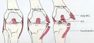
Understanding grade 3 mcl rupture
A Grade 3 MCL rupture represents the most severe form of medial collateral ligament injury, characterized by a complete tear of the ligament. This injury leads to significant instability in the knee joint, making everyday activities and movement challenging. The term “rupture” here signifies a complete break, unlike lesser grades which may involve partial tears.
Definition and Severity
In a Grade 3 MCL rupture, the ligament is entirely torn, resulting in gross laxity and instability in the knee joint8. This severity level is distinct from lower grades, where the ligament may still retain some integrity. The complete tear means the knee may feel unstable, even during simple movements like walking or standing.
Differentiating from Lower Grade Tears
While Grade 1 and Grade 2 MCL injuries involve partial tears with some ligament integrity remaining, a Grade 3 rupture eliminates all functional stability9. This distinction is crucial as it directly impacts treatment approaches. For instance, Grade 1 injuries may heal within 1-3 weeks with minimal intervention, whereas Grade 3 ruptures often require more intensive treatment, possibly including surgery10.
Treatment options for Grade 3 MCL ruptures vary, ranging from non-surgical methods like physical therapy and bracing to surgical intervention in severe cases. Physical therapy plays a significant role in the rehabilitation process, helping restore strength and stability to the knee. A brace may also be recommended to provide additional support during the healing process8.
Understanding the severity of a Grade 3 MCL rupture is essential for developing an effective treatment plan. Whether through conservative management or surgery, the goal is to restore knee stability and function, allowing patients to resume their normal activities without discomfort or limitation.
Common Causes and Risk Factors
Understanding the causes of MCL injuries is crucial for prevention and treatment. These injuries often result from a combination of sports-related trauma, accidents, and repetitive stress. Let’s explore these factors in detail.
Sports-Induced Trauma
Sports, especially those involving pivoting and contact, are a major contributor to MCL injuries. Activities like football, basketball, and skiing often involve sudden movements that can lead to valgus stress on the knee, increasing the risk of an MCL tear11. For instance, a football player making a sharp cut or a skier falling awkwardly can experience this type of stress. These scenarios serve as a test of the ligament’s strength and can reveal its vulnerability, particularly in athletes who frequently engage in high-impact exercises12.

Additionally, repetitive exercise in sports can weaken the MCL over time, making it more susceptible to injury. This is why many athletes experience MCL tears during intense training sessions or competitions2.
Accidents and Repetitive Stress
Accidents, such as falls or direct blows to the knee, can also cause MCL injuries. These incidents often occur during everyday activities or non-sports related events. Furthermore, repetitive stress from activities like running or cycling can gradually strain the ligament, leading to a tear over time11. This type of stress is a common risk factor, especially for individuals who engage in repetitive knee-bending exercises without proper warm-up or rest12.
It’s important to monitor how these injuries manifest week by week during recovery. For example, a minor injury may heal within a few weeks, while a more severe tear could take months to recover from, requiring extensive rehabilitation and exercise routines2.
Recognizing Signs and Symptoms
Identifying the signs and symptoms of a serious knee injury is crucial for prompt treatment and recovery. When it comes to medial collateral ligament injuries, especially severe ones, recognizing the key indicators can make a significant difference in outcomes.
Pain, Swelling, and Instability
The most immediate symptoms of a severe MCL injury often include sharp pain and noticeable swelling around the knee13. This swelling can signal internal damage, as the body reacts to the injury by increasing blood flow to the area. Many athletes report that the pain is intense and debilitating, making it difficult to bear weight or move the knee14.
In addition to pain and swelling, instability is a hallmark symptom of a severe MCL injury. The knee may feel unstable or wobbly, even during simple movements like standing or walking. This instability can be particularly challenging for athletes who rely on their knees for performance15.
Other Warning Signs
Beyond the immediate symptoms, there are other warning signs that may indicate a severe MCL injury. For example, a popping sound at the time of injury can signal a complete tear of the ligament13. Additionally, bruising and limited range of motion are common in more severe cases14.
Key warning signs to watch for:
- Sharp pain and swelling around the knee.
- Instability or a feeling of the knee “giving way.”
- A popping sound at the time of injury.
- Bruising and limited mobility.
Understanding these symptoms and seeking medical attention promptly is essential for an effective recovery. Whether you’re a professional or recreational athlete, recognizing these signs can help prevent further damage and ensure the best possible outcome15.
Diagnostic Methods and Imaging Techniques
Accurate diagnosis is crucial for effective treatment of MCL injuries. Clinicians employ a combination of physical examinations and advanced imaging to assess ligament integrity and determine the severity of the injury.
Physical Examination Procedures
A thorough physical examination is the first step in diagnosing an MCL injury. The valgus stress test, where the knee is bent slightly and pressure is applied to the inside, is commonly used to assess joint stability16. This test helps identify laxity and pain, which are indicators of a potential MCL injury.
Advanced Imaging Modalities
Magnetic resonance imaging (MRI) is the gold standard for diagnosing MCL injuries due to its high accuracy in visualizing soft tissue damage16. MRI provides detailed images of the ligament, helping to confirm the extent of the tear. Other imaging techniques like stress radiography and ultrasonography are also used to evaluate joint stability and ligament integrity.
Stress radiography measures the medial joint space under valgus stress, with a gap difference of 2.0 mm indicating a positive finding17. Ultrasonography offers a cost-effective and real-time alternative, though it may lack the detailed resolution of MRI. Each modality has its strengths, and clinicians often combine them for a comprehensive diagnosis.
Resonance imaging techniques, including MRI, play a vital role in confirming the diagnosis and guiding treatment decisions. While MRI is more accurate, stress radiography and ultrasonography provide valuable complementary information, especially in cases where MRI is unavailable16.
Non-Surgical Treatment and Recovery Options
Non-surgical approaches are often the first line of defense in treating MCL injuries, especially for less severe cases. These methods focus on reducing inflammation, restoring joint function, and strengthening the surrounding muscles. Let’s explore the most effective strategies.
The RICE Protocol
The RICE protocol—Rest, Ice, Compression, and Elevation—is a cornerstone of initial recovery. Rest prevents further injury, while ice reduces swelling and pain18. Compression and elevation further minimize swelling, promoting a conducive environment for healing. Studies show that adhering to the RICE protocol can significantly accelerate recovery, especially in the first 48 hours19.
For instance, in case studies, patients who strictly followed the RICE protocol experienced reduced symptoms and faster return to activity compared to those who did not19. This approach is particularly effective for partial tears, where the ligament still retains some integrity.
Physical Therapy Strategies
A well-structured physical therapy program is essential for regaining strength and stability. Therapists often design exercises to target the muscles around the knee, improving joint function and reducing the risk of future injuries. For example, strengthening the quadriceps and hamstrings can provide additional support to the knee18.
In many documented case examples, patients who underwent consistent therapy saw significant improvements in joint stability and a reduction in symptoms like pain and instability. Tracking these symptoms over time helps tailor the treatment plan, ensuring optimal recovery.
Surgical Treatment Options and Considerations
When non-surgical methods fall short, surgical intervention becomes necessary for certain cases of MCL injuries. This is especially true for severe injuries or those combined with other ligament damage, where surgery can provide the stability needed for proper healing20.
Primary Repair vs. Reconstruction
Primary repair involves stitching the torn ligament back together, while reconstruction replaces the damaged ligament with a graft. Studies show that primary repair has a higher failure rate of 20%, compared to reconstruction’s 4%21. Reconstruction is often recommended for complete tears to restore strength and stability.
Minimally Invasive Techniques
Minimally invasive surgeries are now common, offering less tissue damage and faster recovery. These procedures are typically outpatient, allowing patients to return home the same day20.
| Procedure | Success Rate | Failure Rate | Return to Sports |
|---|---|---|---|
| Primary Repair | 76% | 20% | 9.2 weeks |
| Reconstruction | 98% | 4% | 11.5 weeks |
Magnetic resonance imaging plays a crucial role in surgical planning, providing detailed views of the ligament and surrounding tissues21. This helps surgeons decide the best approach, ensuring optimal outcomes and restoring knee strength effectively.
Rehabilitation and Post-Treatment Exercise Programs
Recovering from an MCL injury requires a structured approach to rehabilitation. A well-designed exercise program is essential to restore strength, motion, and stability in the knee. This section outlines the key components of an effective rehabilitation plan, including strengthening exercises and the use of a knee brace.
Strengthening Exercises
Strengthening exercises are crucial for rebuilding muscle support around the knee. These exercises target the quadriceps, hamstrings, and surrounding muscles to enhance stability and prevent future injuries. For instance, straight leg raises and heel slides are commonly recommended, with repetitions ranging from 10 to 15 times per set22. Additionally, activities like partial squats and wall slides are effective for improving strength without putting excessive strain on the knee23.
Using a Knee Brace
A knee brace can provide additional support during the healing process. It helps control instability and prevents excessive lateral movement, which can exacerbate the injury. The brace is typically worn for 3 to 6 weeks, depending on the severity of the injury22. This supportive measure is particularly beneficial during the gradual return to sport activities, allowing the knee to heal while maintaining functional mobility.
Protocols for resuming sport activities often include a phased approach, starting with low-impact exercises and progressively increasing intensity. Strengthening exercises, combined with proper bracing, play a vital role in restoring knee stability and preventing future injuries. For more detailed guidelines on rehabilitation, refer to the sports medicine protocol.
Expert Insights and Case Examples
Leading professionals in orthopedic sports medicine emphasize the importance of personalized treatment plans for MCL injuries. Experts from institutions like UCSF highlight that recovery outcomes can vary widely, stressing the need for tailored approaches based on the patient’s leg strength and activity level24.
Key insights from professionals include:
- Restoring full range of motion is a critical rehabilitation goal to ensure proper knee function25.
- Case studies demonstrate that treatment outcomes can differ significantly, even for similar injuries24.
- Personalized treatment plans, considering the patient’s leg strength and activity level, lead to better recovery results25.
Experts also recommend regular monitoring of recovery progress to adjust treatment plans as needed. This approach ensures that patients achieve optimal outcomes, whether they are athletes or individuals with less active lifestyles24.
Innovations and Future Research in MCL Injury Management
Emerging therapies and cutting-edge research are revolutionizing the treatment of medial collateral ligament injuries, offering new hope for patients seeking to restore motion and stability. These advancements not only address the femur and surrounding bone structures but also have implications for related injuries, such as those involving the anterior cruciate ligament.
Emerging Therapies
Recent studies have highlighted novel surgical techniques that improve outcomes for the femur area and overall knee structure26. These techniques consider the integrity of surrounding bone, ensuring more effective healing and stability. Additionally, regenerative medicine approaches, such as stem cell therapies, are showing promise in enhancing motion recovery post-treatment27.
These innovations also impact the management of related injuries, such as those to the anterior cruciate ligament. By addressing both ligaments simultaneously, treatments can provide comprehensive care, leading to better long-term outcomes1. The integration of these therapies into clinical practice is expected to significantly improve the recovery process for patients with complex knee injuries.
Conclusion
In conclusion, managing a grade 3 MCL rupture requires a comprehensive approach that addresses every part of the recovery process, from diagnosis to rehabilitation. This injury, involving a complete tear of the medial collateral ligament, is the most severe form of MCL injury and often necessitates specialized care to restore knee stability and function.
The importance of an integrated treatment plan cannot be overstated. Diagnosis typically involves advanced imaging techniques like MRI, which provides detailed views of the ligament and surrounding tissues28. Treatment options range from non-surgical methods, such as physical therapy and bracing, to surgical intervention in severe cases. For instance, studies show that reconstruction of the MCL provides more rotational stability compared to direct repair in combined ACL-MCL injuries29.
Rehabilitation plays a critical role in restoring strength and stability to the knee. Exercises such as heel slides, wall slides, and straight leg raises are commonly recommended to improve range of motion and strength28. The use of a knee brace is also essential for providing additional support during the healing process, especially for Grade 3 injuries, which often require a brace for at least six weeks30.
Research and innovation are key to improving outcomes for patients with complex ligament injuries. Emerging therapies, such as regenerative medicine, are showing promise in enhancing recovery and restoring motion in the knee29. Additionally, advancements in surgical techniques are providing more effective solutions for addressing both cruciate and collateral ligament injuries, ensuring comprehensive care for patients with multi-ligament knee damage.
In summary, every part of the recovery process, from initial diagnosis to long-term rehabilitation, is critical for achieving full function and stability in the knee. By adopting an integrated approach and staying at the forefront of research and innovation, we can ensure the best possible outcomes for patients with grade 3 MCL ruptures.
FAQ
What is a medial collateral ligament (MCL) tear?
An MCL tear is an injury to the medial collateral ligament, a key stabilizer on the inner side of the knee. It can range from mild stretches to complete ruptures, often caused by direct blows or twists to the knee.
What are the symptoms of an MCL injury?
Symptoms include pain on the inner knee, swelling, and instability. In severe cases, there may be a inability to bear weight or a feeling of the knee giving way.
How is an MCL tear diagnosed?
Diagnosis typically involves a physical exam, including tests like the valgus stress test. Imaging such as X-rays or an MRI may be used to confirm the extent of the injury and rule out other issues.
What treatment options are available for an MCL tear?
Treatment depends on the severity. Mild to moderate tears often heal with the RICE protocol (Rest, Ice, Compression, Elevation) and physical therapy. Severe tears may require surgery followed by rehabilitation.
How long does it take to recover from an MCL injury?
Recovery time varies. Mild injuries may heal in a few weeks, while more severe tears can take several months, especially if surgery is involved.
What is the difference between MCL and ACL tears?
The MCL is on the inner knee, while the ACL is in the center. ACL tears often require surgery, while MCL tears can sometimes heal without it.
Can I prevent an MCL injury?
Yes. Strengthening the muscles around the knee, improving flexibility, and using proper techniques during sports can reduce the risk of injury.
Do I need surgery for an MCL tear?
Surgery is usually reserved for severe tears that don’t respond to non-surgical treatments. Most MCL injuries can heal with conservative care.
What role does physical therapy play in recovery?
Physical therapy is crucial for restoring strength, range of motion, and stability. It helps prevent chronic instability and ensures proper healing of the ligament.
How does an MCL tear affect sports activities?
It can sideline athletes temporarily. Proper rehabilitation is essential to safely return to sports, usually once strength and stability are restored.