When dealing with a suspected MCL injury, one of the first questions that comes to mind is whether an MRI is necessary for diagnosis. This is especially crucial for athletes and individuals who lead active lifestyles, as accurate diagnosis is key to proper treatment and recovery.
The MCL, or medial collateral ligament, plays a vital role in stabilizing the knee. Injuries to this ligament often occur due to direct blows to the knee, sudden twists, or awkward landings during sports. Understanding whether an MRI is required can help patients and medical professionals make informed decisions about the next steps in treatment.
While physical examinations and patient history are essential, imaging can provide a clearer picture of the injury’s severity. An MRI offers detailed visuals of soft tissues, helping to confirm the extent of the MCL damage. This can be critical in determining whether surgery is needed or if a conservative approach would be more effective.
At the same time, it’s important to consider the costs and benefits of undergoing an MRI. For some patients, an MRI might not be necessary if a thorough examination and other imaging tests, like an X-ray, have already provided sufficient information. However, in cases where the injury is severe or the diagnosis is unclear, an MRI becomes a valuable tool.
We aim to uncover the facts and provide a clear understanding of when an MRI is necessary for diagnosing an MCL sprain. By exploring the role of MRI in diagnosis, we hope to empower patients and athletes with the knowledge they need to make informed decisions about their care.
Key Takeaways
- Understanding the necessity of MRI for MCL sprain diagnosis is crucial for both patients and athletes.
- The MCL is essential for knee stability, and injuries often occur due to direct trauma or sudden movements.
- Imaging, including MRI, plays a significant role in accurately diagnosing the severity of MCL injuries.
- While MRIs provide detailed images of soft tissues, they may not always be necessary for every patient.
- The decision to use MRI should be based on the severity of symptoms and the clarity of diagnosis from other methods.

Understanding MCL Injuries and Their Impact
The medial collateral ligament (MCL) is a crucial ligament located on the medial (inner) side of the knee. It plays a vital role in stabilizing the knee joint, especially during activities that involve twisting or bending. Injuries to the MCL can significantly impact both daily life and athletic performance.
What is the Medial Collateral Ligament?
The MCL is one of the four major ligaments in the knee, providing stability to the medial (inner) aspect of the joint. It connects the femur (thigh bone) to the tibia (shin bone) and is essential for maintaining proper knee alignment during movement.
How MCL Injuries Affect Daily Life and Sports
MCL injuries can lead to pain, swelling, and instability in the knee. For athletes, this often means a temporary halt in their training or competition. Even non-athletes may find everyday activities like walking or climbing stairs challenging due to the discomfort and instability caused by an MCL injury.
| Ligament | Function | Injury Impact | Common Symptoms | Importance |
|---|---|---|---|---|
| Medial Collateral Ligament (MCL) | Stabilizes the inner aspect of the knee | Causes instability and pain | Pain on the inner side of the knee, swelling, limited mobility | Critical for knee stability during sports and daily activities |
Understanding the types of MCL injuries is crucial for both patients and athletes. Whether it’s a minor sprain or a complete tear, recognizing the symptoms early can lead to more effective treatment and faster recovery.
In sports like football, soccer, and skiing, MCL injuries are common due to the high-impact nature of these activities. However, even non-athletes can suffer from MCL injuries due to everyday accidents, such as slipping on icy sidewalks or twisting the knee awkwardly.
In conclusion, MCL injuries can have a significant impact on both athletic performance and daily life. Understanding the nature of these injuries and their symptoms is the first step toward effective treatment and recovery.
MCL Injury Anatomy and Function
The knee’s medial collateral ligament (MCL) is a vital component of its structural integrity. Located on the inner side of the knee, the MCL connects the femur (thigh bone) to the tibia (shin bone), playing a crucial role in stabilizing the joint during movement.
Anatomical Overview of the Knee’s Medial Structures
The MCL is a tough band of connective tissue that runs from the femur’s medial epicondyle to the tibia’s medial surface. This ligament is essential for preventing excessive inward bending of the knee, a common injury mechanism in sports. The MCL works in tandem with the medial meniscus and surrounding tendons to maintain joint stability and facilitate smooth motion.
Understanding the MCL’s structure is key to grasping how injuries occur. The ligament is composed of collagen fibers arranged in a specific pattern to withstand tensile forces. Its attachment sites on the femur and tibia are critical for its function. Variations in the MCL’s size and attachment points can influence the type and severity of injuries, with some individuals being more prone to certain injury patterns due to their anatomical makeup.
The interplay between ligaments and tendons in the knee is complex. While ligaments like the MCL provide stability, tendons connect muscles to bones, enabling movement. This intricate balance is essential for normal knee function, and any disruption can lead to pain and instability.
Symptoms and Grading of MCL Sprains
Identifying the symptoms and understanding the severity of an MCL injury is crucial for effective treatment. Accurate diagnosis ensures proper care and recovery plans.
Recognizing the Common Symptoms
- Pain: Often felt on the inner side of the knee.
- Swelling: Develops within hours of injury.
- Instability: Feeling of the knee giving way.
- Popping Sensation: Heard or felt during injury.
These symptoms can significantly impact daily activities and sports participation, causing discomfort and limited mobility.
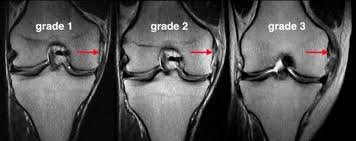
Understanding Injury Severity: Grade 1, 2, and 3
- Grade 1: Mild injury with minimal pain and no instability.
- Grade 2: Partial tear with some laxity and moderate pain.
- Grade 3: Complete tear causing severe instability and pain.
Physical examinations and stress tests help determine the grade, guiding treatment decisions.

Recognizing these symptoms early leads to better outcomes and faster recovery.
Imaging Techniques for Knee Injuries
Accurate diagnosis of knee injuries often requires the use of imaging techniques. These methods help healthcare providers assess the extent of damage to ligaments, bones, and other tissues. Among the most common imaging techniques are X-rays, stress radiography, and magnetic resonance imaging (MRI). Each has its unique benefits and limitations.
X-ray and Stress Radiography Explained
X-rays are often the first imaging test used for knee injuries. They are particularly useful for identifying bone fractures or misalignments. X-rays work by sending low-dose radiation through the body to create images of internal structures. In the context of MCL injuries, X-rays can help rule out fractures or bone-related issues.
Stress radiography takes X-ray imaging a step further. This technique involves applying gentle pressure to the knee to assess joint stability. By measuring the joint space, healthcare providers can identify ligament damage. For example, stress radiography can reveal joint space widening, which may indicate an MCL injury.
Magnetic Resonance Imaging: How It Works
Magnetic resonance imaging (MRI) is a powerful diagnostic tool that provides detailed images of soft tissues, including ligaments, tendons, and cartilage. MRI uses magnetic fields and radio waves to create high-resolution images of the knee. This non-invasive procedure is especially valuable for diagnosing MCL injuries, as it can reveal partial or complete ligament tears.
One of the key advantages of MRI is its high accuracy, with studies showing it can diagnose MCL injuries with up to 90% accuracy. Additionally, MRI can detect associated injuries, such as meniscal tears or bone bruises, which might not be visible on X-rays.
| Imaging Method | Key Features | Benefits | Limitations |
|---|---|---|---|
| X-ray | Uses radiation to image bones | Quick, cost-effective, detects bone fractures | Does not show soft tissue damage |
| Stress Radiography | Applies pressure to assess joint stability | Identifies joint space widening and ligament damage | May cause discomfort, limited to joint alignment |
| MRI | High-resolution images of soft tissues | Accurate diagnosis of ligament and tendon injuries | Higher cost, longer procedure time |
For patients undergoing these imaging procedures, the experience is relatively straightforward. X-rays and stress radiography are quick, while MRI requires lying still in a machine for about 30-60 minutes. Understanding these options helps patients and healthcare providers choose the most appropriate test for their situation.
For more detailed insights into the role of MRI in diagnosing knee injuries, refer to this study on advanced imaging techniques.
is mri needed for mcl sprain diagnosis
When evaluating a suspected MCL injury, a key question arises: Is an MRI necessary for diagnosis? This is especially important for athletes and active individuals seeking precise treatment plans.
While physical exams are essential, they may not always provide a complete picture. MRI scans become invaluable in scenarios where physical exam findings are unclear or when additional injuries, such as an ACL tear, are suspected. MRI offers detailed images of soft tissues, helping to identify partial or complete ligament tears that other tests might miss.
In comparing diagnostic tools, X-rays are quick and cost-effective but lack the ability to show soft tissue damage. Stress radiography can assess joint stability but may cause discomfort. MRI, while more expensive, provides high-resolution images of ligaments and tendons, making it the most accurate method for diagnosing severe injuries.
Timing is crucial in MRI use. It’s most beneficial after initial assessments when more severe injuries are suspected. Early use can prevent prolonged recovery and ensure proper treatment. For example, if a patient experiences persistent instability despite conservative treatment, an MRI can reveal underlying issues guiding further interventions.
We use these tests to confirm the extent of ligament damage and develop personalized treatment plans. Detailed imaging ensures that each patient receives care tailored to their specific injury, whether through rehabilitation or surgery.
Conservative Treatments and Rehabilitation Options
For many patients, recovering from an MCL injury doesn’t require surgery. Conservative treatments and rehabilitation strategies can effectively promote healing and restore knee function. These approaches are tailored to the injury’s severity and the patient’s lifestyle, ensuring a personalized recovery plan.
Rest, Ice, and Bracing Strategies
The foundation of conservative treatment is the RICE method: Rest, Ice, Compression, and Elevation. Resting the knee is crucial to avoid further injury, while ice helps reduce swelling. Compression, often achieved with an elastic bandage or sleeve, supports the knee and limits swelling. Elevation, keeping the leg above heart level, also aids in reducing swelling.
Knee braces are another key component. These braces restrict unwanted movement, providing stability and protection during the healing process. For example, an MCL brace can prevent excessive stress on the injured ligament, allowing it to heal properly. Patients often wear braces for several weeks, depending on the injury’s severity.
The Role of Physical Therapy in Recovery
Physical therapy plays a vital role in restoring strength and mobility. A structured rehab program typically begins with gentle exercises to maintain range of motion and progresses to strengthening workouts. Techniques like neuromuscular reeducation help improve balance and coordination, reducing the risk of future injuries.
Therapists may incorporate exercises such as straight leg raises, hamstring curls, and quadriceps sets. These exercises target the muscles around the knee, enhancing stability without putting excessive strain on the MCL. Over time, more dynamic exercises and agility drills are introduced to prepare the patient for return to normal activities.
| Treatment Method | Features | Benefits | Limitations |
|---|---|---|---|
| Rest, Ice, Compression, Elevation (RICE) | Initial care for acute injuries | Reduces swelling, promotes healing | Requires patience and adherence |
| Knee Bracing | Provides structural support | Stabilizes the knee, prevents re-injury | May feel restrictive |
| Physical Therapy | Structured exercises, neuromuscular training | Restores strength, mobility, and function | Requires consistent participation |
Recovery timelines vary by injury grade. Grade 1 injuries may heal within 1-3 weeks, while Grade 2 injuries could take 4-6 weeks. Grade 3 injuries, involving a complete tear, may require 8-12 weeks of conservative treatment before considering surgery. Monitoring symptoms and swelling is essential, as improvements often indicate progress.
These strategies not only reduce pain but also prevent further injury. By combining rest, bracing, and therapy, patients can achieve a full recovery without surgical intervention. Tailoring treatment to the injury type ensures the best outcomes for each patient.
Surgical Interventions for Severe MCL Injuries
In cases where conservative treatments fail to heal an MCL injury, surgical intervention becomes necessary. Surgery is often recommended for complete tears or when other ligaments, like the ACL, are also damaged.
When Surgery Becomes Necessary
Surgery is typically reserved for severe MCL tears that don’t respond to non-surgical treatments. A surgeon may recommend surgery if the injury causes significant instability or if there are additional injuries, such as an ACL tear.
Recovery and Post-Surgical Care
After surgery, patients use crutches and wear a brace to protect the knee. Physical therapy begins shortly after surgery to restore strength and mobility. Recovery time varies, but most patients can return to normal activities within 6 to 9 months.
| Surgical Procedure | Description | Recovery Time |
|---|---|---|
| Ligament Repair | Fixing the torn ligament | 6-9 months |
| Ligament Reconstruction | Using a tendon graft | 9-12 months |
Comparison of MRI with Other Diagnostic Tools
When evaluating knee injuries, choosing the right diagnostic tool is essential for accurate results. While several imaging methods are available, MRI stands out for its detailed insights, especially in cases involving ligament injuries.
Advantages of Using MRI for MCL Injuries
- Higher Accuracy: MRI provides up to 90% accuracy in diagnosing MCL tears, surpassing X-rays and stress radiographs which focus on bones and joint alignment.
- Detection of Associated Injuries: MRI can identify additional issues like ACL tears or meniscal damage, offering a comprehensive view of the injury.
- Influence on Treatment Plans: Detailed MRI results guide personalized treatment, whether through conservative methods or surgery, ensuring targeted care.
For instance, in a case where an athlete suffered persistent instability, an MRI revealed a complete MCL tear and an ACL injury, necessitating surgical intervention. This highlights MRI’s role in preventing misdiagnosis and tailoring treatments effectively.
While X-rays and stress tests are useful, they lack MRI’s ability to detail soft tissue damage. MRI’s clarity is unmatched, making it indispensable for complex injuries. It ensures precise diagnosis and recovery planning, crucial for both athletes and non-athletes alike.
Prevention Strategies and Long-Term Recovery
Preventing future MCL injuries and maintaining knee health is essential for both athletes and non-athletes. By adopting proper strength conditioning and rehabilitation practices, individuals can reduce the risk of re-injury and promote long-term joint stability.
Injury Prevention and Strength Conditioning
A strong foundation is key to preventing MCL injuries. Engaging in regular strength training exercises, such as squats and lunges, can enhance the muscles around the knee, providing additional support to the ligaments. Proper warm-up and cool-down routines are also crucial, as they prepare the muscles for activity and aid in recovery.
- Strengthen the surrounding muscles through targeted exercises.
- Include balance and agility drills to improve overall knee stability.
- Wear appropriate footwear to reduce the risk of awkward movements.
Maintaining Knee Health After Recovery
Even after recovery, it’s important to continue preventative exercises. Regular physical therapy sessions can help maintain joint stability and prevent future injuries. Monitoring knee health over time and scheduling routine check-ups with a healthcare provider are also vital for long-term recovery.
| Preventative Measure | Benefits |
|---|---|
| Strength Training | Enhances muscle support around the knee |
| Proper Warm-Up | Reduces injury risk during activities |
| Regular Therapy | Maintains joint stability and function |
By prioritizing these strategies, patients can effectively protect their ligaments and enjoy lasting knee health.
Conclusion
Accurate diagnosis and effective treatment of MCL injuries are essential for restoring knee function and preventing future issues. Whether through advanced imaging like resonance-based scans or physical examinations, understanding the injury’s severity is crucial. This knowledge guides personalized treatment plans, which may include conservative approaches like bracing and therapy or surgical interventions for severe cases.
Understanding the structure and symptoms of MCL injuries, along with their grading, helps in making informed decisions. From non-invasive methods to surgery, each treatment option should be tailored to the patient’s specific needs. Additionally, adopting prevention strategies such as strength training and proper warm-ups can significantly reduce the risk of re-injury.
Our commitment is to guide patients through every part of their recovery journey. We encourage consulting with a qualified surgeon or specialist to ensure the best possible outcomes. If you’re dealing with an MCL injury, don’t hesitate to seek further advice and take proactive steps toward a full recovery.
FAQ
What is the medial collateral ligament (MCL)?
The medial collateral ligament (MCL) is a vital structure in the knee that provides stability to the inner (medial) aspect of the joint. It connects the femur (thigh bone) to the tibia (shin bone) and plays a crucial role in preventing excessive movement during activities like walking or sports.
What are the common symptoms of an MCL injury?
Common symptoms of an MCL injury include pain on the inner side of the knee, swelling, and a feeling of instability. In more severe cases, you may hear a popping sound at the time of injury, followed by difficulty bearing weight or moving the knee.
How is an MCL injury typically diagnosed?
Diagnosis of an MCL injury often involves a combination of a physical exam and imaging tests. Your doctor may perform a valgus stress test to assess ligament integrity. While X-rays can rule out fractures, magnetic resonance imaging (MRI) is the gold standard for confirming the extent of ligament and surrounding tissue damage.
Do all MCL injuries require surgery?
No, most MCL injuries do not require surgery. Partial tears and less severe injuries (Grade 1 or 2) typically heal well with conservative treatments like bracing, physical therapy, and rest. Surgery is usually reserved for complete tears (Grade 3) or cases where the injury occurs alongside other ligament damage, such as an ACL tear.
How long does it take to recover from an MCL injury?
Recovery time for an MCL injury varies depending on the severity. Grade 1 injuries may heal within 1-2 weeks, while Grade 2 injuries could take 2-4 weeks. More severe Grade 3 injuries might require 4-6 weeks or longer, especially if surgery is involved. Proper rehabilitation is essential to restore strength and prevent future injuries.
Can I return to sports after an MCL injury?
Yes, many people return to sports after recovering from an MCL injury. However, it’s important to wait until your knee has fully healed and you’ve regained strength and stability. Rushing back too soon can increase the risk of re-injury. Your healthcare provider will help determine when it’s safe to resume athletic activities.
How can I prevent an MCL injury?
Preventing an MCL injury involves strengthening the muscles around the knee through targeted exercises, improving flexibility, and ensuring proper technique during sports. Wearing appropriate protective gear and avoiding overuse can also reduce the risk of injury.