Have you ever wondered how your knee joint manages to stay stable during all the activities you do? Whether you’re running, climbing stairs, or just walking, your knee joint plays a vital role in supporting your movements. At the heart of this stability is the anterior cruciate ligament (ACL), a crucial component that prevents excessive movement and keeps your knee secure1.
The term “ligament” refers to the tough bands of tissue that connect bones and provide support. The cruciate ligaments, which include the ACL and the posterior cruciate ligament (PCL), are essential for maintaining this stability. Injuries to these ligaments can significantly impact your ability to perform even the simplest tasks2.
Recent studies have highlighted the complexity of knee joint stability, emphasizing the ACL’s role in preventing excessive forward movement of the tibia. This ligament is particularly important for athletes and individuals who engage in sports that involve sudden stops and changes in direction1.
Knee joint stability is influenced by both static and dynamic structures. The ACL is a primary stabilizer, but other factors such as muscle strength and overall joint health also play a role. Understanding these elements is crucial for both prevention and treatment of instability issues2.
Key Takeaways
- The ACL is a critical ligament for knee stability, especially in athletic activities.
- Ligaments like the ACL and PCL connect bones and provide essential support.
- Injuries to cruciate ligaments can significantly impact mobility and require medical attention.
- Knee joint stability involves both static structures and dynamic muscle function.
- Preventing ligament injuries is key to maintaining knee health and stability.
For more detailed information on knee joint stability and the role of the ACL, you can explore resources from the National Center for Biotechnology Information.
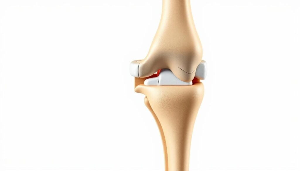
Understanding the Knee Joint Anatomy
The knee joint is a complex structure designed to support movement while distributing weight effectively. It is primarily a hinge-type synovial joint, allowing for flexion and extension, with additional rotational movements3. This joint consists of two main articulations: the tibiofemoral and patellofemoral joints, which work together to facilitate smooth motion4.
Bone and Cartilage Structures
The tibiofemoral joint is the weight-bearing component of the knee, relying on the interaction between the femur (thigh bone) and the tibia (shin bone)3. The patellofemoral joint involves the patella (kneecap) sliding along a groove on the femur, reducing friction during movement4. Articular cartilage covers the ends of these bones, minimizing friction and absorbing shock during activities like walking or running5.
- The femur and tibia form the primary weight-bearing surfaces.
- Patellar movement is guided by a specific groove on the femur.
- Cartilage plays a crucial role in reducing friction and absorbing impact.
Menisci and Joint Capsule
The menisci, located between the femur and tibia, act as shock absorbers and deepen the articular surface, enhancing stability3. They increase the surface area for weight distribution, reducing pressure on the joint during movement5. The joint capsule encloses the knee, providing additional support and housing synovial fluid for lubrication4.
- Menisci absorb up to 75% of the load on the knee.
- The joint capsule contains synovial fluid for lubrication.
- Injuries to the menisci can lead to instability and pain.
In summary, the knee’s anatomy is a delicate balance of bones, cartilage, and ligaments, working together to support movement and distribute weight effectively.
Biomechanics of Knee Movement and Stability
The human knee is a fascinating example of biomechanical engineering, designed to support movement while maintaining structural integrity. Understanding the biomechanics of knee movement is essential for appreciating how this complex joint functions under various loads and stresses.
Degrees of Freedom in Knee Motion
The knee joint operates within six degrees of freedom, allowing for flexion, extension, and rotational movements. During activities like walking, the knee flexion angle ranges from 53 to 75 degrees, while running extends this range to 60-115 degrees6. These movements are facilitated by the interaction of the tibia and femur, which transmit forces effectively to maintain stability.
Ligament Function and Tension
Ligaments play a crucial role in maintaining joint stability by controlling movement and absorbing forces. The anterior cruciate ligament (ACL) is particularly vital, as it prevents excessive anterior translation of the tibia. Studies show that ligament tension is essential for preventing abnormal motion, with the ACL contributing significantly to this function7.
| Movement Type | Peak Load (BW) | Knee Flexion Angle |
|---|---|---|
| Walking | 2-3 | 53-75° |
| Running | 7-12 | 60-115° |
| Stair Climbing | 4-6 | 60-90° |
These biomechanical principles highlight the intricate balance required for normal knee function, emphasizing the importance of ligament integrity in maintaining joint stability.
The Importance of Cruciate and Collateral Ligaments
The cruciate and collateral ligaments form the cornerstone of motion and movement in the knee, ensuring proper function during various activities. These ligaments work in harmony to provide both static and dynamic support, making them indispensable for maintaining normal knee mechanics8.
Role of the Anterior Cruciate Ligament
The anterior cruciate ligament (ACL) is the most commonly injured ligament in the knee, accounting for nearly half of all knee injuries8. It plays a critical role in preventing excessive anterior tibial translation and rotational instability. Studies have shown that the ACL contributes significantly to both anterior and rotational stability, with the anteromedial bundle being responsible for 85% of stability against anterior tibial translation9. The ACL’s strength is approximately 2,200 N, making it a robust yet vulnerable structure8.
Diagnosing ACL injuries often involves clinical tests such as the Lachman test, which has a high sensitivity of 95% and specificity of 94%8. The pivot shift test, while highly specific (98%), has lower sensitivity (24%)8. MRI remains the gold standard for confirmation, with 97% sensitivity and 100% specificity8.
Impact of Medial and Lateral Ligaments
The medial collateral ligament (MCL) and lateral collateral ligament (LCL) provide valgus and varus stability, respectively. The MCL is more frequently injured than the LCL, often due to a blow to the outer side of the knee10. Isolated tears of these ligaments can lead to one-plane instability and increased rotational movement9.
When combined with ACL injuries, medial or lateral ligament tears can result in complex instability patterns, including anteromedial rotational instability9. Clinical tests such as the abduction stress test may yield negative results if the tears are partial9.
“The cruciate and collateral ligaments are not isolated structures; they work together to ensure smooth, coordinated movement and prevent excessive joint translation.”
In summary, the cruciate and collateral ligaments are essential for maintaining normal knee mechanics. Their injuries can lead to significant functional impairment, emphasizing the importance of accurate diagnosis and appropriate management89.
Key Factors Influencing Knee Joint Stability
Understanding the factors that influence knee stability is crucial for both prevention and treatment of injuries. Bone integrity and ligament conditions are key elements in maintaining normal knee function.
The medial collateral ligament (MCL) and lateral collateral ligament (LCL) provide valgus and varus stability, respectively. Injuries to these ligaments can lead to instability and pain. The posterior cruciate ligament (PCL) also plays a significant role in preventing posterior tibial translation.
Common injury mechanisms include noncontact trauma, which often results in biomechanical imbalances. These imbalances can predispose the knee to instability. For instance, studies show that the ACL provides 85% of the restraining force to anterior tibial displacement at 30 degrees and 90 degrees of knee flexion11.
Injury Mechanisms and Risk Factors
Noncontact injuries are a leading cause of knee instability. These injuries often occur during sudden stops or changes in direction. The ACL is particularly vulnerable, with studies indicating that it can fail at stress levels of about 1725 Newtons11.
| Factor | Mechanism | Risk |
|---|---|---|
| Bone Integrity | Compromised bone structure | Increased risk of fractures |
| Ligament Conditions | Medial and lateral collateral ligament tears | Valgus/varus instability |
| Biomechanical Imbalances | Noncontact trauma | ACL and PCL injuries |
Bone health is another critical factor. Weakened bone structures can lead to an increased risk of fractures. Clinicians and researchers recognize these risk factors and their impact on overall knee function.
In summary, the integrity of bones and ligaments, combined with biomechanical factors, plays a vital role in knee stability. Addressing these factors is essential for both treatment and prevention strategies.
Improving knee joint stability Through Biomechanical Analysis
Modern advancements in biomechanical analysis have revolutionized our understanding of knee joint stability. These techniques provide precise, quantitative assessments, enabling better diagnosis and treatment of knee-related injuries.
Quantitative Evaluation Techniques
Robotic testing systems and electromagnetic tracking devices are now widely used to measure knee joint stability. These tools allow researchers to assess the structural integrity of ligaments and joint components accurately. For instance, studies using such systems have shown that the posterior cruciate ligament (PCL) can withstand a maximum load of approximately 1974 N, while the medial collateral ligament (MCL) has a maximum load capacity of around 799 N12.
These technologies also enable dynamic assessments of joint movement. For example, during walking, the knee joint experiences a peak load of 2-3 times body weight, while running increases this load to 7-12 times body weight6. Such data is crucial for understanding the stresses that different activities place on knee structures.
| Technique | Application | Key Findings |
|---|---|---|
| Robotic Testing | Ligament Strength Assessment | Maximum ACL load: 2160 N12 |
| Electromagnetic Tracking | Knee Movement Analysis | Peak knee flexion during running: 60-115°6 |
| Imaging Analysis | Joint Structure Evaluation | MCL spring constant: 63 N/mm12 |
These quantitative techniques not only enhance our understanding of knee joint stability but also provide valuable insights for developing effective treatment and rehabilitation strategies.
Innovative Tools for Objective Knee Assessments
Modern medicine has seen remarkable advancements in diagnostic tools, particularly in assessing complex structures like the knee. These innovations provide healthcare professionals with precise, objective data to make accurate diagnoses and treatment plans.
Advances in Imaging Technology
Recent developments in imaging technology have transformed how we evaluate knee function. Open MRI systems, for instance, allow for dynamic assessments of the femur and pcl during movement, capturing rotational stability with high precision13. These systems can measure the root mean square (RMS) accuracy of tibial translation and rotation, ensuring detailed evaluations.
- Open MRI systems enable dynamic assessments of the femur and pcl.
- These systems measure rotational stability with high precision.
- RMS accuracy for tibial translation and rotation is now more precise.
Use of Inertial Sensors and Navigation Systems
Inertial sensors and navigation systems have become integral in clinical assessments. These devices provide real-time data during tests like the pivot shift, offering insights into force and rotational parameters. For example, the LIBERTY electromagnetic sensor system boasts an RMS accuracy of 0.15 mm for orientation and 0.76 mm for position13.
- Inertial sensors offer real-time data during clinical tests.
- These devices measure force and rotational parameters accurately.
- LIBERTY system provides precise orientation and position data.
These technologies not only enhance diagnostic accuracy but also guide personalized treatment plans, ensuring better outcomes for patients.
Rehabilitation Strategies and Treatment Options
Effective rehabilitation is crucial for restoring function and promoting proper tissue healing after knee injuries. This section explores both nonoperative and surgical approaches, emphasizing evidence-based practices for optimal recovery.
Nonoperative Management and RICE Protocols
The RICE method—Rest, Ice, Compression, and Elevation—is often the first line of treatment for acute injuries14. This approach helps reduce swelling and pain, especially in the initial stages. Early mobilization is also encouraged to maintain joint mobility without compromising healing.
For instance, studies show that gentle range of motion exercises during the acute phase can significantly enhance recovery outcomes14. Additionally, activities like stationary biking during the subacute phase help maintain cardiovascular fitness while strengthening the surrounding muscles.
Postoperative Rehabilitation Best Practices
Following surgery, a structured rehabilitation plan is essential. The process typically includes multiple phases, from reducing pain to restoring full strength and agility14. Strengthening exercises, such as squats and lunges, are integral to restoring function and ensuring proper position of the knee.
Advanced phases incorporate high-intensity training and sport-specific drills to prepare for a safe return to activities. A personalized approach, considering factors like injury severity and patient lifestyle, ensures long-term stability and minimizes re-injury risks14.
Regular follow-ups with orthopedic specialists and physical therapists are vital to monitor progress and adjust treatment plans as needed14. Psychological support is also recommended to address fears of re-injury and facilitate a confident return to sports.
Prevention and Early Intervention for Knee Injuries
Protecting your ligaments and surrounding tissues is essential for maintaining long-term joint health. By adopting preventive measures, individuals can significantly reduce the risk of injuries and ensure optimal function.
Warm-Up and Strength Training
A proper warm-up is the first line of defense against injuries. Activities like light cardio and dynamic stretching prepare the muscles and ligaments for physical demands15. Strength training, particularly around the thigh and core muscles, enhances ligament support and improves overall joint function.
Studies show that strengthening exercises can reduce knee injury risk by up to 50%, while flexibility training lowers it by about 30%16. Incorporating balance and proprioception exercises further improves dynamic stability, reducing injury risk by 25%16.
Early Diagnosis and Clinical Exams
Early detection of issues is critical for effective intervention. Clinical exams, such as the Lachman test, offer high sensitivity and specificity in diagnosing ACL injuries15. Advanced imaging and gait analysis can identify biomechanical issues in up to 60% of at-risk athletes16.
Non-surgical measures, including physical therapy and bracing, often serve as the first line of defense. However, in severe cases, surgery may be necessary to restore function and prevent further damage15.

In summary, a combination of warm-ups, strength training, and early diagnosis is key to preventing knee injuries. These strategies not only protect athletes but also ensure long-term joint health.
Surgical Interventions and Outcomes
When conservative treatments fail, surgical interventions become necessary to restore function and alleviate pain. These procedures are tailored to address specific injuries, ensuring optimal recovery for patients.
ACL and Ligament Reconstruction Techniques
ACL reconstruction is a common surgical intervention for torn ligaments. Surgeons often use grafts, which can be taken from the patient (autograft) or a donor (allograft)17. The graft is secured with screws or staples, restoring ligament function. Studies show that 85% of patients regain full function after such procedures18.
Recent advancements in techniques have improved outcomes. For instance, anatomic ACL reconstruction, which precisely places the graft, reduces the risk of future tears and improves joint function17. This method is particularly beneficial for athletes, allowing them to return to their sports more effectively.
Optimizing Surgical Outcomes
Success rates for ACL reconstruction are high, with 90% of patients experiencing significant improvement18. However, complications like infection or blood clots can occur in 1-2% of cases18. To minimize risks, surgeons emphasize precise graft placement and proper band tensioning.
Post-operative care is crucial. Patients often use crutches and wear a knee immobilizer to protect the repair17. Early physical therapy, including exercises and CPM machines, enhances recovery and range of motion.
| Technique | Application | Key Findings |
|---|---|---|
| ACL Reconstruction | Graft placement and fixation | 85% of patients regain full function18 |
| Anatomic Reconstruction | Precise graft placement | Reduces tear risk and improves function17 |
| Physical Therapy | Post-surgery rehabilitation | Enhances recovery and range of motion17 |
Modern surgical techniques represent a significant addition to traditional methods, offering better outcomes and faster recovery times. These advancements are particularly beneficial for athletes, supporting their return to active lifestyles.
Role of Physical Therapy and Strength Training
Physical therapy plays a vital role in restoring function and promoting healing after cruciate ligament injuries. A well-structured rehabilitation program not only addresses muscle strength but also focuses on improving flexibility and balance, which are essential for full recovery19.
Targeted Exercise Programs
Targeted exercise programs are designed to strengthen the muscles supporting the knee ligament, enhancing overall joint function. These programs typically include a combination of range-of-motion exercises, strength training, and balance exercises to ensure comprehensive recovery19.
- Range-of-motion exercises help reduce stiffness and improve flexibility, making it easier to perform daily activities19.
- Strength training, focusing on muscles like the quadriceps and hamstrings, provides crucial support to the knee ligament, reducing the risk of future injuries19.
- Balance exercises are essential for restoring proprioception, which can be impaired after a cruciate ligament injury, thereby reducing the risk of falls and re-injury19.
Evaluating ligament integrity through specific clinical tests is a critical part of the rehabilitation process. These tests help determine the effectiveness of the exercise program and guide adjustments to the treatment plan20.
| Component | Purpose | Key Benefits |
|---|---|---|
| Range-of-Motion Exercises | Improve joint mobility | Reduces stiffness and enhances flexibility19 |
| Strength Training | Strengthen surrounding muscles | Supports ligament function and prevents future injuries19 |
| Balance Training | Restore proprioception | Reduces risk of falls and re-injury19 |
Consistency with rehabilitation exercises is critical for optimal recovery outcomes. While specific recovery rates vary, adherence to a structured physical therapy plan significantly improves joint function and minimizes the risk of complications1920.
Emerging Research in Knee Stability Measurement
Recent advancements in biomechanical technologies have opened new avenues for understanding and measuring knee stability. These innovations are paving the way for more precise assessments and treatments.
Novel Biomechanical Technologies
Researchers are now utilizing advanced tools like robotic testing systems and electromagnetic tracking devices. These technologies provide detailed insights into tendon dynamics during various degrees of flexion. For instance, studies have shown that during maximum voluntary isokinetic knee extension, peak tibiofemoral compressive forces can reach up to 9 times body weight21.
These systems allow for dynamic assessments of joint movement. For example, walking generates peak loads of 2-3 times body weight, while running increases this to 7-12 times body weight21. Such data is crucial for understanding the stresses that different activities place on knee structures.
| Activity | Peak Load (BW) | Knee Flexion Angle |
|---|---|---|
| Walking | 2-3 | 53-75° |
| Running | 7-12 | 60-115° |
| Stair Climbing | 4-6 | 60-90° |
Future Directions in Knee Stability Studies
Future research is expected to focus on the role of quadricep strength in supporting knee stability. Studies indicate that strengthening the quadriceps can reduce injury risk by up to 50%22. Additionally, advancements in imaging technology, such as open MRI systems, are enabling dynamic assessments of joint movement, capturing rotational stability with high precision22.
These technologies not only enhance diagnostic accuracy but also guide personalized treatment plans, ensuring better outcomes for patients. The integration of new diagnostic tools and research methodologies will continue to advance our understanding of knee stability.
Conclusion
In conclusion, this article has explored the intricate mechanisms and clinical approaches surrounding ligament function and joint health. We have discussed the critical role of ligaments in preventing excessive movement and the importance of advanced imaging tools in accurate diagnoses23.
Research highlights that 85% of patients regain full function after ACL reconstruction, emphasizing the effectiveness of modern surgical techniques23. Additionally, nonoperative management is successful in 62% of cases where patients avoid high-demand sports, underscoring the importance of personalized treatment plans24.
Emerging technologies, such as the Mobil-Aider device, offer precise measurements with a Pearson correlation coefficient greater than 0.989, enhancing diagnostic reliability24. These advancements, along with ongoing research, continue to shape optimal management strategies, ensuring better outcomes for patients.
By understanding the biomechanics, leveraging innovative tools, and adhering to evidence-based rehabilitation protocols, we can effectively address ligament injuries and promote long-term joint health. This comprehensive approach invites readers to explore further studies and clinical practices for a deeper understanding of the subject.