Have you ever wondered how your knees support your every move, twist, and turn? The answer lies in the complex yet fascinating anatomy of the knee ligaments. These fibrous bands play a crucial role in providing stability and strength to the joint, enabling us to walk, run, and jump without a second thought.
At the heart of the knee’s structure are four major ligaments: the anterior cruciate ligament (ACL), posterior cruciate ligament (PCL), medial collateral ligament (MCL), and lateral collateral ligament (LCL). The ACL, measuring 33mm in length and 11mm in width, is one of the most commonly injured ligaments, particularly in high-impact sports like football and skiing1. In contrast, the PCL, slightly larger at 38mm by 13mm, often sustains injuries from direct impacts, such as those occurring in car accidents1.
The MCL, with a tensile strength of 4000 N, is the strongest of the four and is more frequently injured than the LCL21. Understanding the anatomy and functions of these ligaments is essential for preventing injuries and evaluating treatment options, which can range from conservative approaches like physical therapy to surgical interventions1.
Key Takeaways
- The knee contains four major ligaments: ACL, PCL, MCL, and LCL.
- The ACL is one of the most commonly injured ligaments, especially in sports.
- The MCL is stronger than the LCL and is more frequently injured.
- Ligament injuries can result from sudden impacts or twists.
- Treatment options vary from conservative methods to surgery, depending on severity.
For more detailed information on knee ligament injuries, visit Hopkins Medicine.
Introduction to the Knee Joint and Its Ligaments
The knee joint, the largest and most pivotal joint in the body, is essential for supporting daily movement. It functions primarily as a hinge joint, allowing leg flexion and extension while providing stability through its complex structure34.
The Role of Ligaments in Knee Stability
Ligaments act as elastic bands, providing crucial stability to the knee. The anterior cruciate ligament (ACL) and posterior cruciate ligament (PCL) are vital for preventing excessive forward and backward movement of the femur, respectively45. The medial collateral ligament (MCL) stabilizes the inner knee, often working in tandem with the joint capsule5.
Why a Detailed Understanding Matters
Understanding the knee’s structure is key to preventing injuries and developing effective treatments. For instance, knowing how the MCL and ACL function can help athletes and individuals adopt preventive measures, reducing the risk of injuries like the “unhappy triad”5. This knowledge also aids in recognizing early symptoms, such as instability or pain, which are crucial for timely intervention.
Anatomical Overview of the Knee
The knee is a fascinating joint that combines strength and flexibility to support our daily activities. Its complex structure includes both bony and soft tissue components, each playing a vital role in movement and stability.
Bone Structures and Articulating Surfaces
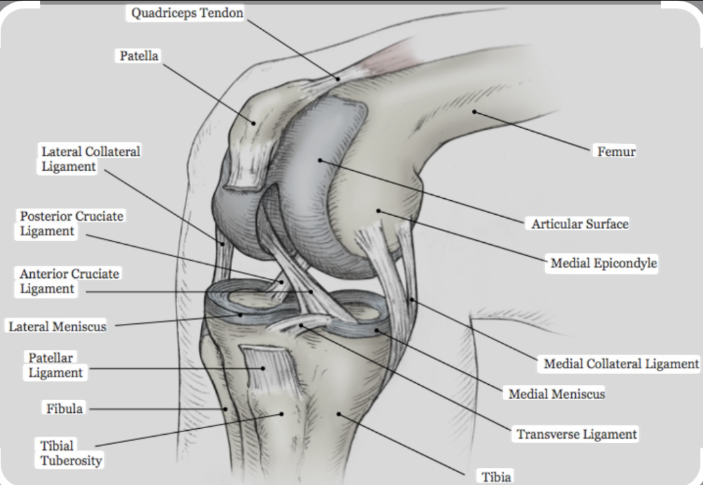
The knee joint is formed by three main bones: the femur (thigh bone), tibia (shin bone), and patella (kneecap)67. These bones come together to create the tibiofemoral and patellofemoral joints. The femur’s rounded condyles fit into the tibia’s flat surface, allowing for smooth movement, while the patella glides in a groove on the femur, enhancing leg extension7.
The joint can bear loads of up to 4.9 times body weight, making it incredibly resilient during activities like walking or running8.
Cartilage, Menisci, and Bursae Explained
Hyaline cartilage covers the ends of the bones, reducing friction and acting as a shock absorber8. The menisci, located between the femur and tibia, are “C”-shaped cartilage structures that absorb up to 75% of the knee’s load, preventing bone-on-bone contact8.
Bursae, small fluid-filled sacs, cushion the joint, reducing friction between soft tissues and bones. The suprapatellar and infrapatellar bursae are key examples, facilitating smooth knee flexion and extension8.
These structures work together to ensure smooth, pain-free movement, highlighting the knee’s intricate design for both stability and mobility.
knee ligament anatomy: Structure and Components
The knee joint’s stability is largely attributed to its intricate ligament system, which includes both cruciate and collateral ligaments. These components work in harmony to provide structural support and enable smooth movement.
Cruciate Ligaments: ACL and PCL Functions
The anterior cruciate ligament (ACL) and posterior cruciate ligament (PCL) are central to controlling the tibia’s movement relative to the femur8. The ACL prevents excessive forward movement of the tibia, while the PCL stabilizes it from moving too far backward. Together, they form a cross within the knee, hence the name “cruciate ligaments.”
Collateral Ligaments: MCL and LCL Insights
The medial collateral ligament (MCL) and lateral collateral ligament (LCL) provide side-to-side stability. The MCL, stronger than the LCL, is more prone to injury and plays a crucial role in stabilizing the inner knee7. The LCL supports the outer aspect, working alongside the joint capsule to maintain alignment during movement.
| Ligament | Attachment | Function |
|---|---|---|
| ACL | Femur to Tibia | Prevents tibia from sliding forward |
| PCL | Femur to Tibia | Prevents tibia from sliding backward |
| MCL | Femur to Tibia (inner) | Stabilizes inner knee |
| LCL | Femur to Fibula (outer) | Stabilizes outer knee |
Understanding these ligaments’ roles is vital for diagnosing injuries and developing effective treatments. Injuries to these structures can significantly impact joint stability and function, often requiring specialized care for recovery.
Mechanics and Movement of the Knee Joint
The knee joint is a remarkable mechanism designed for both strength and agility. It supports our every step, twist, and turn with precision. Understanding its mechanics is key to appreciating how it enables such a wide range of movements.
Flexion, Extension, and Rotational Dynamics
The knee primarily moves in two directions: flexion (bending) and extension (straightening). During flexion, the knee can bend up to 160°, allowing activities like squatting or sitting9. Extension brings the knee back to its straight position, essential for standing and walking.
In addition to these primary movements, the knee also allows for medial (inward) and lateral (outward) rotation. When the knee is bent, it can rotate inward by about 10° and outward by up to 30°, enhancing mobility during activities like pivoting or changing direction9.
How Ligaments Support Motion
Ligaments play a crucial role in stabilizing the knee during these movements. The ACL and PCL prevent excessive forward and backward movement, while the MCL and LCL provide side-to-side stability. This ligamentous support ensures that the knee moves smoothly and remains stable.
The interplay between muscles and ligaments is essential for controlled motion. Muscles generate the force needed for movement, while ligaments guide and limit the joint’s movement, preventing overextension or unnatural twists. This teamwork allows for precise and efficient motion, whether you’re walking or running.
Proper biomechanical function also protects the knee from overuse injuries. When the knee moves as it should, it distributes forces evenly, reducing stress on individual structures like ligaments and cartilage. This natural distribution is why maintaining good movement mechanics is vital for injury prevention.
In everyday activities and sports, understanding these mechanics can enhance performance and reduce injury risk. By appreciating how the knee moves and the role of ligaments, we can better protect this essential joint.
Common Knee Ligament Injuries
Knee ligament injuries are a common challenge for both athletes and individuals performing everyday activities. These injuries often result from sudden twists, direct impacts, or overuse, leading to significant discomfort and impaired mobility.
Injury Mechanisms in Sports and Daily Life
Sports like football, basketball, and skiing pose a high risk of ligament injuries due to their dynamic movements. Sudden stops or changes in direction can strain the anterior cruciate ligament (ACL), making it the most frequently injured ligament1011.
Medial collateral ligament (MCL) injuries often occur from direct blows to the knee’s inner side, commonly seen in contact sports. Conversely, posterior cruciate ligament (PCL) injuries typically result from direct impacts to the front of the bent knee, such as in car accidents1011.
Recognizing Symptoms and Early Warning Signs
Immediate symptoms of a ligament injury may include a popping sound, knee buckling, and rapid swelling. These signs indicate potential damage and necessitate prompt medical evaluation to prevent further harm1011.
Early recognition is crucial for effective treatment. Consulting a healthcare professional promptly can lead to better outcomes, whether through conservative methods or surgery, depending on the injury’s severity11.
Incorporating strengthening exercises and proper techniques in daily activities can significantly reduce injury risks. Awareness and preventive measures are key to maintaining knee health and functionality.
Diagnostic Techniques for Knee Ligament Injuries
Accurate diagnosis is crucial for effectively treating knee ligament injuries. Modern diagnostic methods combine clinical exams with advanced imaging to ensure precise assessments.
Imaging Methods: X-Ray, MRI, and Arthroscopy
X-rays are often the first step in diagnosing knee injuries. They help rule out bone fractures and assess joint alignment1213. While X-rays provide valuable information, they don’t show soft tissues like ligaments, making them insufficient for comprehensive diagnosis.
MRI scans offer detailed images of ligaments, muscles, and surrounding soft tissues. They are particularly effective in identifying ACL and PCL injuries, with sensitivities of 86% and 95% respectively1314. This makes MRI the primary diagnostic tool for ligament injuries.
Arthroscopy is a minimally invasive procedure that allows direct visualization of the joint. It not only helps diagnose injuries but can also enable immediate treatment in some cases14. Arthroscopy is especially useful for confirming ACL tears, with a sensitivity of 92-100%13.
Importance of Accurate Diagnosis
An accurate diagnosis guides appropriate treatment plans, whether through conservative methods or surgery. Early and precise detection of injuries like ACL or PCL tears can prevent further damage and improve outcomes1214.
Understanding what to expect during these procedures helps patients feel more prepared. From the non-invasive nature of MRI to the surgical aspects of arthroscopy, clear communication ensures informed decisions and better recovery paths.

Treatment and Rehabilitation Approaches
Treating knee ligament injuries requires a personalized approach, blending non-surgical interventions with surgical options when necessary. The goal is to restore joint stability, strength, and mobility while minimizing recovery time.
Non-Surgical Interventions and Rehabilitation Exercises
For mild to moderate injuries, non-surgical treatments are often effective. These include rest, anti-inflammatory medications, and targeted rehabilitation exercises1516. Physical therapy plays a crucial role in strengthening the muscles around the knee, improving stability, and restoring range of motion.
Surgical Options and Recovery Strategies
Surgery is typically recommended for severe ligament tears, especially when other treatments fail to provide relief. Common procedures include ligament repair or reconstruction. Recovery often involves several months of rehabilitation, with patients using crutches and knee immobilizers initially1517.
Prevention Tips from Sports Medicine Experts
Preventing injuries is as important as treatment. Strengthening exercises, proper warm-ups, and using appropriate techniques can significantly reduce injury risks. Programs like those offered by UH Sports Medicine highlight the importance of prehab and neuromuscular training17.
| Treatment Type | Description | Recovery Time |
|---|---|---|
| Non-Surgical | Rest, medication, physical therapy | Varies, 2-6 weeks |
| Surgical | Ligament repair/reconstruction | 6-12 months |
| Rehabilitation | Exercise programs, physiotherapy | 3-6 months |
Collaboration between patients and healthcare providers is key to determining the best treatment plan. Whether through non-invasive methods or surgery, the focus remains on restoring function and preventing future injuries.
Impact of Sports and Lifestyle on Knee Health
Sports and lifestyle significantly influence knee health, affecting both injury risks and long-term joint stability. Regular physical activity, while beneficial, can sometimes predispose the knee to injuries, especially when proper precautions are not taken.
How Physical Activity Influences Injury Risks
Participating in sports like football and basketball can increase the risk of ligament injuries, with ACL injuries accounting for approximately 40% of all sports-related knee injuries18. Additionally, gaining just five kilograms of excess weight can negatively impact knee health, particularly for individuals with pre-existing conditions like arthritis19.
Lifestyle factors such as weight management and overall fitness also play a crucial role in knee stability. Sedentary lifestyles can lead to a loss of muscle tone, which may result in knee pain or instability during physical activities19.
| Sport | Common Injuries | Prevention Tips |
|---|---|---|
| Football | ACL tears, meniscal injuries | Proper warm-ups, strength training |
| Basketball | PCL injuries, patellar tendinitis | Dynamic stretching, balanced landing techniques |
| Running | Meniscal tears, IT band syndrome | Gradual mileage increase, proper footwear |
Engaging in sports requires a balance between maintaining an active lifestyle and protecting vulnerable knee structures. Strengthening exercises and proper techniques can significantly reduce injury risks, as highlighted by sports medicine experts18.
For more detailed insights on preventing sports-related knee injuries, visit this resource.
Conclusion
In conclusion, understanding the intricate anatomy of the knee joint and its ligaments is essential for maintaining mobility and preventing injuries. The knee, formed by the femur, tibia, and patella, relies on four primary ligaments—the ACL, PCL, MCL, and LCL—to provide stability and support during movement2021.
Proper biomechanics and timely diagnosis are critical for effective treatment. Advanced imaging techniques like MRI and arthroscopy, as detailed by sources such as Johns Hopkins Medicine and Cleveland Clinic, play a vital role in accurately diagnosing injuries, ensuring appropriate treatment plans2221. Understanding the ligaments’ functions and the joint’s mechanics can guide preventive measures and rehabilitation strategies, enhancing long-term knee health.
Modern treatment approaches, ranging from non-surgical interventions to surgical reconstruction, are tailored to restore joint stability and function. By adopting preventive strategies, such as strengthening exercises and proper techniques, individuals can significantly reduce injury risks2021.
If you experience any knee discomfort, consulting a healthcare professional is crucial for early intervention and effective management. Proactive care and awareness are key to maintaining knee wellness and ensuring continued mobility and strength.