Have you ever wondered why some knee injuries linger despite months of rest and therapy? While conservative treatments work for many, certain ligament damage requires precision-focused solutions to restore full mobility. At our practice, we prioritize understanding your unique needs before recommending any course of action.
Knee stability relies heavily on the medial collateral ligament (MCL), which often bears the brunt of sports injuries or accidents. When this critical structure becomes compromised, everyday activities like climbing stairs or walking can feel impossible. Our team specializes in evaluating whether surgical intervention truly offers the best path forward – or if targeted rehabilitation could achieve similar results.
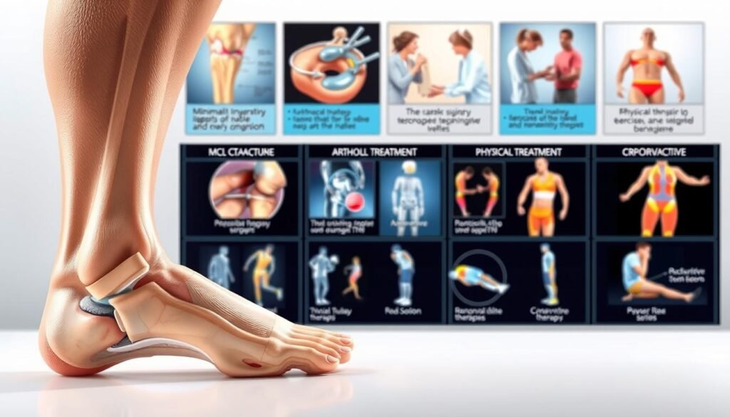
We combine cutting-edge diagnostic tools with decades of hands-on experience to create personalized recovery plans. Through minimally invasive approaches, we aim to reduce downtime while maximizing long-term joint function. Every decision focuses on one goal: helping you regain confidence in your body’s capabilities without unnecessary procedures.
Key Takeaways
- Specialized evaluation determines if surgical solutions are essential for ligament recovery
- Advanced techniques prioritize knee stability and accelerated healing timelines
- Personalized care plans address both immediate discomfort and future joint resilience
- Minimally invasive methods minimize tissue disruption during procedures
- Transparent decision-making process empowers patients throughout treatment
Overview of the Medial Collateral Ligament and Knee Anatomy
Among the key players in knee stability, one ligament stands out for its crucial role in preventing inward collapse. The medial collateral ligament (MCL) spans from the femur to the tibia’s upper section, forming a thick band along the inner knee. This structure acts as a primary stabilizer, resisting excessive side-to-side motion while permitting natural rotation.

Function of the Medial Collateral Ligament
We educate patients about this ligament’s dual purpose: maintaining joint integrity during movement and absorbing lateral forces. Its position between the thighbone and shinbone creates a protective barrier against inward leg displacement. Simultaneously, it works with surrounding tissues to enable smooth bending and twisting motions essential for daily activities.
Anatomical Relationship within the Knee Joint
The MCL’s broad structure connects to muscles and supporting tissues, forming a collaborative network. Forces applied to the knee’s outer side often stress this inner stabilizer, explaining common injury patterns. Understanding these connections helps us explain why targeted treatments preserve overall joint function beyond just repairing isolated damage.
Understanding MCL Injuries and Overall Knee Health
What makes certain activities more likely to harm knee stability? Protecting joint health starts with recognizing how daily movements and unexpected impacts affect vulnerable structures. We focus on educating patients about prevention strategies while addressing existing damage through evidence-based methods.
Common Causes of MCL Injuries
Lateral force to the outer knee remains the primary culprit behind most ligament damage. Collisions during football tackles or hockey checks often create this stress pattern. Even non-contact events like awkward landings after jumps can overstretch tissues beyond their limits.
Risk Factors for Knee Ligament Damage
High-intensity sports dominate our injury risk assessments, but we also evaluate workplace habits and fitness routines. Occupations requiring repetitive kneeling or heavy lifting gradually weaken stabilizing structures. Weekend warriors face particular challenges when sudden activity spikes meet underprepared muscles.
| Activity/Event | Potential Impact |
|---|---|
| Side collisions in contact sports | Forces knee inward, stressing medial ligaments |
| Quick pivots in basketball/soccer | Twists joint beyond natural rotation range |
| Falls on icy surfaces | Unexpected leg splaying strains inner knee |
We prioritize teaching proper warm-up techniques and strengthening exercises tailored to each patient’s lifestyle. Early intervention often prevents minor sprains from becoming chronic issues affecting mobility.
Recognizing the Symptoms of an MCL Tear
How can you tell if your knee injury involves ligament damage? Symptoms often appear suddenly and intensify within hours. Inner knee discomfort paired with joint instability typically signals potential damage to critical stabilizing tissues.

Identifying Key Warning Signs
We guide patients through recognizing early indicators. A sharp “pop” sensation along the inner leg during impact frequently marks the injury moment. Tenderness concentrates where the ligament connects thigh and shin bones.
Swelling develops gradually, often peaking 6-12 hours post-injury. Many describe their knee feeling unreliable during weight-bearing activities, like climbing stairs. Bruising patterns help us assess whether other structures sustained damage.
| Symptom | Significance |
|---|---|
| Audible popping sound | Suggests sudden ligament stress |
| Localized tenderness | Pinpoints injury location |
| Progressive swelling | Indicates inflammatory response |
| Weight-bearing difficulty | Reveals functional limitations |
| Locking sensations | Warns of potential complications |
Stiffness often follows as the body restricts movement to protect healing tissues. We differentiate between normal recovery responses and signs requiring urgent care. Joint catching or locking suggests possible cartilage involvement needing advanced imaging.
Diagnosing MCL Injuries Effectively
Accurate identification of joint damage begins with thorough detective work. Our team pieces together clues from your experience and advanced tools to map out recovery pathways. This method eliminates guesswork while respecting your body’s unique healing capacity.
Building the Full Picture
We start by reconstructing your injury’s story. When did discomfort begin? Did you hear a pop during impact? Previous joint issues receive equal attention, as past events often influence current treatment strategies. Gentle pressure tests follow, assessing tenderness along the inner knee.
Two positioning techniques reveal ligament stability:
- Leg straightened to evaluate overall joint integrity
- Knee bent 30 degrees to isolate specific structures
Seeing Beneath the Surface
While many cases resolve through physical exams alone, we deploy imaging when questions remain. Standard X-rays rule out fractures, while MRI scans offer detailed soft tissue views. Stress X-rays prove particularly useful, capturing joint space changes under controlled pressure.
Our diagnostic approach balances efficiency with precision:
| Method | Key Benefit |
|---|---|
| Physical Exam | Immediate stability assessment |
| MRI Scan | 90% accuracy for ligament evaluation |
| Stress Imaging | Reveals hidden joint laxity |
We translate complex medical information into clear action plans. Every finding gets explained using visual aids and plain language, empowering you to make informed decisions about next steps.
Treatment Options for MCL Injuries
Choosing the right path to recovery requires a clear assessment of your injury. Many ligament issues respond well to structured rehabilitation programs thanks to the body’s natural healing capacity. We focus on methods that align with your lifestyle while protecting joint function.
Non-Surgical Management and Rehabilitation
Our team designs progressive therapy plans starting with gentle range-of-motion exercises. These evolve into strength-building routines targeting muscles around the joint. Neuromuscular reeducation helps retrain movement patterns disrupted by injury.
Functional training mimics real-world activities like squatting or pivoting. This approach rebuilds stability while reducing re-injury risks. Regular progress evaluations ensure your plan adapts as healing advances.
Surgical Interventions and When to Consider Them
We reserve operative solutions for complex cases involving multiple damaged structures. Severe instability or failed conservative care may indicate the need for reconstruction. Our surgeons use tissue-sparing techniques to preserve healthy anatomy.
Decisions always balance potential benefits against recovery timelines. Post-procedure rehabilitation remains critical for restoring full mobility. Every plan prioritizes returning you to activities safely and efficiently.
MCL Pain Surgery: When is Surgery Necessary?
While many ligament injuries heal without procedures, some scenarios require precise solutions. We determine if surgical approaches will improve long-term stability by evaluating activity demands, coexisting damage, and recovery objectives.
Prioritizing Joint Resilience Through Specialized Care
Combined injuries often dictate our recommendations. When ACL damage accompanies ligament tears, non-surgical methods may fail to address complex instability. Athletes frequently benefit from these interventions due to extreme joint stress during competition.
Persistent instability after rehabilitation signals potential need for advanced solutions. We assess tear grade and location alongside other tissue involvement. Our plans balance anatomical repair with strategies to support future physical demands, ensuring confidence in every movement.
Decisions always center on restoring functional capacity while minimizing downtime. Whether addressing high-grade injuries or sport-specific needs, we tailor solutions that align with your body’s mechanics and goals.