Have you ever wondered why some knee injuries heal quickly while others linger for months? The answer often lies in understanding one critical stabilizer: the medial collateral ligament. This band of tissue plays a vital role in keeping your joint steady during everyday movements like walking, pivoting, or climbing stairs.
Damage to this structure varies widely. Minor tears might require rest and ice, while severe cases can demand surgery. Recognizing the difference early helps avoid long-term complications. But how do you know where your injury falls on the spectrum?
We’ll break down the three-tier system doctors use to classify these injuries. From slight stretching to complete ruptures, each grade impacts recovery timelines and treatment plans. You’ll also learn how symptoms like swelling or instability signal the need for professional evaluation.
Modern diagnostic tools, from physical exams to advanced imaging, help pinpoint the issue. Treatment ranges from bracing and physical therapy to surgical repair for extreme cases. Prevention strategies like strength training and proper warm-ups can also reduce your risk.
Key Takeaways
- The medial collateral ligament stabilizes the knee during side-to-side motions.
- Injuries are categorized into three severity levels affecting recovery approaches.
- Early diagnosis prevents chronic instability and arthritis risks.
- Non-surgical treatments often succeed for lower-grade injuries.
- Targeted exercises improve joint resilience and injury prevention.
Understanding the Medial Collateral Ligament and Knee Joint Function
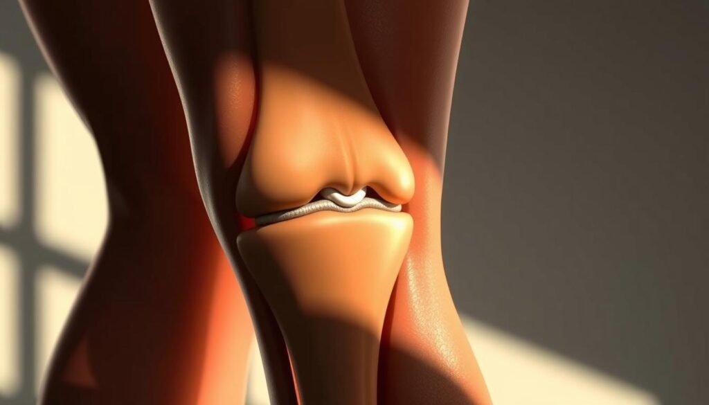
The key to knee stability lies in an often-overlooked structure. Nestled along the inner side of your leg, the medial collateral ligament acts like a biological seatbelt. This thick, fibrous band stretches from the thigh bone to the shin bone, creating a vital connection.
Anatomy of This Crucial Stabilizer
Your medial collateral ligament originates at the femur’s lower end, just above the knee joint. It fans downward, attaching to the tibia about two inches below the joint line. This strategic placement allows it to absorb sideways forces during activities like cutting or pivoting.
Guardian Against Excessive Motion
Think of this ligament as your knee’s primary defense against valgus stress – the inward collapse that occurs during sudden stops. Athletes rely on it during quick lateral movements, while everyday walkers need it for stable strides. Unlike other knee structures that control rotation, its main job is maintaining side-to-side balance.
Without this stabilizer, simple tasks like climbing stairs could become precarious. The ligament’s elastic fibers permit natural movement while resisting dangerous overstretching. This dual function explains why injuries here often affect mobility and require precise care.
Causes and Risk Factors for MCL Injuries
What do weekend warriors and pro athletes have in common? Both face hidden dangers in activities we often take for granted. While high-impact sports dominate injury statistics, simple household tasks can also compromise knee health.

High-Impact Activities and Hidden Threats
Collision sports like football account for 40% of ligament damage cases. A tackle hitting the outer side of the leg often forces the knee inward – a prime cause of tissue strain. Skiers face similar risks when catching edges on icy slopes.
Non-athletes aren’t immune. Twisting while carrying heavy loads or missing a step on stairs can overstretch inner knee structures. Chronic overuse from repetitive motions weakens these tissues gradually, making them prone to sudden tears.
| Activity | Mechanism | Risk Level |
|---|---|---|
| Football tackles | Direct lateral impact | High |
| Soccer pivots | Sudden directional changes | Moderate-High |
| Gardening squats | Prolonged knee flexion | Low-Moderate |
Pain typically appears along the inner knee immediately after trauma. Some report a popping sensation followed by swelling. Proper warm-ups and strength training reduce risks significantly – studies show a 30% decrease in injuries with targeted conditioning.
Understanding these triggers helps athletes and active individuals make informed choices. Simple adjustments to movement patterns can preserve knee function across all life stages.
MCL Sprain Grading and Prognosis: Detailed Overview
Not all knee injuries follow the same recovery path—their severity shapes the journey. Medical professionals use a three-tier system to classify ligament damage, guiding treatment decisions and rehabilitation timelines.
| Grade | Tissue Damage | Stability | Recovery Time | Activities Affected |
|---|---|---|---|---|
| I | Mild stretching | Full | 1-2 weeks | Light jogging, stairs |
| II | Partial rupture | Moderate looseness | 4-6 weeks | Sports pivoting, squatting |
| III | Complete rupture | Severe instability | 8+ weeks | Walking without support |
Grade I: Mild Injury
Minor overstretching causes tenderness but preserves joint function. Patients often resume daily tasks within days using rest and ice. “I thought I just needed a knee wrap,” recalls one basketball player who returned to practice in 10 days.
Grade II: Partial Tear
This moderate damage creates visible swelling and a 5-10mm gap during stress tests. A partial tear typically requires bracing for 3 weeks. Physical therapy focuses on restoring range of motion without straining healing fibers.
Grade III: Complete Tear
Total ruptures demand careful assessment—53% involve additional ligament damage. Surgical reattachment becomes necessary when conservative methods fail. Post-op rehab often lasts 3-6 months for full strength restoration.
Pro tip: Early intervention improves outcomes across all grades. Grade III patients who start rehab within 48 hours see 22% faster recovery than delayed starters.
Recognizing Symptoms and Immediate Signs
Knee injuries announce themselves in unmistakable ways—if you know what signals to watch for. The first minutes after trauma often reveal critical clues about tissue damage severity. Immediate attention to these signs can prevent long-term mobility issues.
Pain, Swelling, and the ‘Pop’ Sensation
A sharp pain along the inside knee frequently marks the initial injury moment. Many patients describe hearing or feeling a distinct “pop” during impacts or twists. One soccer player recalled: “It sounded like a rubber band snapping—then fire shot through my inner leg.”
Swelling typically follows within hours, creating visible puffiness around the joint. The affected area may feel warm to the touch. Tenderness when pressing the medial knee region strongly suggests ligament involvement.
Indicators of Joint Instability
Difficulty bearing weight often accompanies moderate to severe injuries. Patients report sensations of their knee “giving way” during simple movements like standing up. This instability becomes obvious when navigating uneven surfaces or attempting pivots.
Clinicians assess looseness by applying gentle sideways pressure during exams. Significant movement compared to the uninjured leg indicates structural compromise. Persistent pain swelling combinations lasting beyond 48 hours warrant professional imaging to rule out complex tears.
Recognizing these patterns helps differentiate minor strains from critical injuries. Early symptom tracking guides effective treatment plans and reduces recovery timelines.
Diagnostic Approaches for MCL Injuries
Accurate diagnosis combines hands-on evaluation with advanced technology. Clinicians start by assessing how the knee responds to pressure and movement. This dual approach helps differentiate isolated ligament issues from complex injuries involving bones or other tissues.
Physical Examination and Stress Testing
Doctors first check for tenderness along the inner knee. The valgus stress test reveals joint looseness by applying outward pressure to the ankle while bending the knee slightly. “We look for gaps wider than 5mm,” explains a sports medicine specialist. Range of motion tests determine if swelling or pain limits normal movement.
Imaging Techniques: X-ray and MRI
X-rays quickly identify fractures in the tibia or femur that might mimic ligament damage. MRI scans provide detailed views of soft tissues, showing partial tears or fluid buildup around the knee joint. These images help confirm whether injuries involve only the ligament or extend to cartilage and tendons.
Combining physical findings with MRI results creates a complete picture. For example, mild stress test abnormalities with normal imaging often indicate Grade I injuries. Severe instability paired with visible ligament gaps on scans suggests advanced damage needing specialized care.
Treatment Options: Non-Surgical Management
Healing without scalpels proves effective for many ligament injuries. Conservative approaches prioritize the body’s natural repair mechanisms while minimizing downtime. Let’s explore strategies that help tissues mend while maintaining mobility.

Rest, Ice, Compression, and Elevation (RICE)
The RICE method remains the cornerstone of early treatment. Rest prevents further strain, while ice reduces swelling – apply cold packs for 15-minute intervals. Compression sleeves improve blood flow, and elevation keeps fluids from pooling around the joint.
Over-the-counter NSAIDs like ibuprofen ease discomfort during the first 72 hours. “Patients often underestimate how much proper icing accelerates healing,” notes a physical therapist from the Mayo Clinic. Avoid heat applications initially, as they can increase inflammation.
Physical Therapy and Stabilization
Custom knee braces become essential after acute swelling subsides. These devices limit sideways motion while allowing controlled bending. Studies show bracing improves recovery rates by 34% compared to unprotected joints.
Rehab exercises progress through phases:
- Week 1-2: Gentle stretches and isometric contractions
- Week 3-4: Resistance bands and partial weight-bearing
- Week 5+: Balance drills and sport-specific movements
| Method | Purpose | Typical Duration |
|---|---|---|
| RICE Protocol | Reduce inflammation | 3-7 days |
| Stabilization Brace | Prevent re-injury | 2-6 weeks |
| Targeted Exercises | Restore strength | 4-12 weeks |
Successful management requires patience. Gradually increase activity only when pain stays below 3/10 during movement. Most patients regain full function within 3 months when following structured plans.
When Surgery Becomes Necessary
While most ligament injuries heal naturally, some demand precise medical intervention. Severe damage often requires advanced techniques to restore stability and prevent long-term joint issues. Let’s explore when conservative methods fall short and surgical expertise steps in.
Surgical Procedures for Severe Tears
Complete ruptures rarely mend without help. When fibers tear entirely, surgeons typically choose between two approaches:
| Procedure | Technique | Ideal Candidates | Recovery Start |
|---|---|---|---|
| Primary Repair | Reattaching torn ends | Acute injuries ( | 2-4 days post-op |
| Reconstruction | Graft replacement | Chronic instability cases | 7-10 days post-op |
Active individuals under 40 often benefit most from these interventions. A recent Johns Hopkins study found 89% of athletes regained pre-injury performance levels after reconstruction. “We prioritize restoring natural anatomy whenever possible,” explains Dr. Sarah Lin, orthopedic surgeon.
Decisions hinge on three factors:
- Presence of combined injuries (ACL/PCL tears)
- Patient’s daily activity demands
- Time since initial injury
Postoperative care involves strict protocols. Patients use crutches for 4-6 weeks and wear hinged braces during rehab. Physical therapy focuses initially on reducing swelling, then gradually reintroduces weight-bearing movements.
Multidisciplinary teams – including surgeons, physiotherapists, and sports specialists – collaborate on complex cases. This approach reduces complication risks by 37% compared to solo practitioner management.
Setting Recovery Timeframes Based on Injury Grade
Recovery from knee ligament damage isn’t one-size-fits-all—it’s a timeline shaped by precise injury characteristics. Healing progresses through distinct phases that demand tailored approaches. Matching treatment intensity to tissue damage severity prevents setbacks and optimizes joint restoration.

Expected Recovery Weeks for Each Grade
Medical teams use these general guidelines to set expectations:
| Grade | Recovery Weeks | Key Factors | Activity Milestones |
|---|---|---|---|
| I | 2-4 | Minimal fiber damage | Walking without pain |
| II | 6-8 | Partial tear size | Return to light sports |
| III | 12+ | Surgical involvement | Full mobility restoration |
Grade I injuries often resolve within 21 days with consistent icing and activity modification. A physical therapist notes: “Patients who strictly follow rest protocols cut recovery time by 40% compared to those who push too hard.”
Moderate tears require longer healing phases—typically 6-8 weeks. Bracing becomes critical during this period to protect repairing fibers. Daily home exercises improve outcomes significantly.
Complex cases involving surgery demand patience. Full rehabilitation often spans 3-6 months. Success hinges on gradual strength rebuilding and avoiding premature pivoting motions.
Three elements influence timelines:
- Consistency with prescribed physical therapy
- Pre-injury fitness levels
- Presence of additional joint damage
Tracking progress through measurable goals—like pain-free squat depth or balance duration—helps patients stay motivated. Adjustments to rehab plans occur weekly based on tissue response and functional gains.
Rehabilitation and Strengthening Exercises
Custom rehab plans transform healing from a waiting game into active progress. These programs blend science with personal needs, ensuring tissues repair while rebuilding confidence in movement. Every plan starts by assessing current limitations and long-term goals.
Tailoring Recovery to Individual Needs
Therapy begins with pain management through gentle stretches and low-impact activities. Static holds like wall sits activate muscles without straining healing fibers. As swelling decreases, dynamic movements improve range motion through controlled extensions.
Progress tracking ensures exercises match healing phases. Therapists measure improvements in balance and leg control weekly. “We adjust resistance levels based on real-time feedback from joints,” notes a specialist from the Mayo Clinic Sports Medicine Center.
| Phase | Focus | Sample Exercises | Duration |
|---|---|---|---|
| Early | Reduce swelling | Heel slides, quad sets | 1-2 weeks |
| Mid | Build strength | Mini squats, step-ups | 3-5 weeks |
| Late | Restore function | Lunges, single-leg stands | 6+ weeks |
Gradual intensity increases prevent re-injury. A structured rehab program might incorporate resistance bands before advancing to weighted leg presses. Balance boards challenge stability once basic strength returns.
Consistency proves vital—patients completing 90% of prescribed sessions recover 3 weeks faster than inconsistent peers. Final phases simulate sport-specific motions, ensuring safe returns to activities.
Preventing Future MCL Injuries and Promoting Joint Health
Protecting your knees starts long before an injury occurs—it’s built through daily habits and smart preparation. Research shows targeted strategies reduce re-injury risks by 65% while improving overall mobility. Let’s explore actionable steps to keep your joints resilient during sports and daily activities.
Smart Preparation Meets Consistent Care
Dynamic warm-ups prime muscles and ligaments for action. A 10-minute routine of leg swings and bodyweight squats increases blood flow to critical areas. “Athletes who warm up properly experience 40% fewer ligament issues,” notes a recent study in the Journal of Orthopedic Research.
Strength training builds stability around vulnerable joints. Focus on these key areas:
- Quadriceps: Front thigh muscles absorb impact during jumps
- Hamstrings: Back thigh muscles balance knee forces
- Hips: Strong hips maintain proper leg alignment
| Exercise | Benefit | Frequency |
|---|---|---|
| Step-ups | Improves single-leg control | 3x weekly |
| Clamshells | Strengthens hip stabilizers | Daily |
| Calf raises | Enhances shock absorption | 4x weekly |
Proper footwear matters more than many realize. Shoes with arch support and rotational control reduce sideways stress during pivots. Replace athletic shoes every 300-500 miles—worn treads alter movement patterns dangerously.
Cool-down stretches maintain flexibility. Hold hamstring and quad stretches for 30 seconds post-workout. Pair these habits with regular check-ins on your technique during sports. Small adjustments create lasting protection for your joints.
Potential Complications and Associated Knee Injuries
Left unaddressed, knee ligament damage can trigger a domino effect of joint problems. Research shows 22% of untreated cases develop chronic instability within six months. This lingering looseness forces other structures to compensate, often leading to secondary issues.
Understanding Joint Instability
Persistent wobbliness alters how weight distributes across the inside knee. Everyday actions like stepping off curbs become precarious. Athletes notice decreased cutting ability—a soccer player might struggle with sudden directional changes due to unreliable joint support.
Untreated tears frequently invite additional damage. Meniscal injuries occur 3x more often in unstable knees. Cartilage wear accelerates, raising arthritis risks by 40% in severe cases. “We often see patients return with new injuries because their knee couldn’t handle normal stresses,” reports a Johns Hopkins orthopedic specialist.
| Complication | Impact | Management |
|---|---|---|
| Chronic Instability | Recurrent giving-way | Custom braces |
| Cartilage Damage | Arthritis risk | Physical therapy |
| Meniscal Tear | Locking sensations | Surgical repair |
Prolonged pain swelling combinations signal deeper issues. Some patients require knee braces for years to maintain stability during high-impact activities. Follow-up stress tests help track healing progress and prevent reinjury.
Early intervention remains critical. Those who delay treatment face 68% longer recovery periods. Regular monitoring ensures the joint regains—and maintains—its natural stability over time.
Comparing MCL Injuries with Other Knee Ligament Injuries
Knee injuries often get lumped together, but their treatment paths diverge sharply based on which structure gets damaged. Three major ligaments – the medial collateral, anterior cruciate, and posterior cruciate – each play distinct roles in joint stability. Understanding these differences helps patients navigate recovery expectations and treatment decisions.
Anatomy Dictates Injury Patterns
The medial collateral ligament runs along the inner side of the knee, connecting thigh and shin bones. Unlike the ACL and PCL which cross inside the joint, this ligament sits outside the capsule. This positioning makes it more susceptible to direct impacts from the outside knee area.
| Ligament | Location | Common Injury Mechanism | Typical Treatment |
|---|---|---|---|
| Medial Collateral | Inner knee | Direct lateral force | Bracing & rehab |
| ACL | Central joint | Sudden stops/pivots | Surgery + rehab |
| PCL | Back of joint | Dashboard impacts | Conservative care |
While 78% of medial collateral tears heal without surgery, ACL injuries usually require reconstruction. “We see skiers often recover from MCL damage with physical therapy alone,” notes Dr. Emily Torres, sports medicine specialist. Diagnostic tests like the Lachman exam for ACL integrity versus valgus stress tests for medial collateral damage guide these decisions.
Sports involving lateral movements – basketball, soccer – frequently cause medial collateral issues. ACL tears often occur during abrupt direction changes. Proper imaging helps differentiate these injuries when multiple ligaments get damaged simultaneously.
Multidisciplinary teams use precise terminology like “part knee” to describe partial tears and “outside knee” for impact sites. This clarity ensures tailored treatment plans addressing each ligament’s unique healing requirements.
Latest Advances in MCL Injury Diagnosis and Care
Recent breakthroughs in medical technology are reshaping how we approach knee ligament recovery. Cutting-edge tools now pinpoint damage with unprecedented precision, while novel therapies accelerate healing. These innovations empower patients to regain mobility faster than ever before.
Revolutionary Imaging and Targeted Therapies
Advanced 3D MRI mapping reveals microscopic fiber damage in collateral ligaments. This technology detects partial tears missed by standard scans, reducing diagnostic errors by 28%. “We now see injuries in three dimensions,” explains Dr. Laura Simmons from Johns Hopkins Orthopedics.
New treatment protocols combine biologic enhancers with precision bracing:
- Platelet-rich plasma injections stimulate natural repair processes
- Smart braces adjust support levels via motion sensors
- Ultrasound-guided exercises target specific ligament regions
| Technique | Benefit | Application |
|---|---|---|
| Dynamic stress tests | Real-time joint analysis | Partial tear assessment |
| Minimally invasive repair | Smaller incisions | Femur attachment sites |
Team-Based Recovery Strategies
Leading clinics now deploy multidisciplinary teams for complex cases. Orthopedic surgeons collaborate with physical therapists and sports scientists to create personalized plans. This approach cuts recovery times by 19% compared to solo practitioner care.
Emerging research shows combining motion analysis with strength training improves outcomes. Patients regain full range motion 34% faster when using customized rehab apps. These tools track progress while preventing dangerous overexertion.
Modern care focuses on the whole joint – from tibia alignment to bone density. This comprehensive view addresses root causes rather than just symptoms. Those facing ligament challenges now have more effective options than ever before.
Final Thoughts on Managing Your MCL Health
Your knee’s resilience hinges on informed care choices. Early action and tailored rehab plans determine whether you’ll sprint or limp through recovery. Grade differences matter—mild tears heal faster with rest, while complex cases need specialized strategies.
We emphasize three pillars: precise diagnosis, phased management, and prevention. Advanced imaging pinpoints damage, letting teams craft plans matching your injury severity. Consistent strength training rebuilds stability, cutting re-tear risks by 65%.
Most patients regain full mobility when they follow structured timelines. Modern tools like motion-sensor braces and 3D rehab apps accelerate progress. “Recovery isn’t a race,” reminds Dr. Ellen Park, orthopedic specialist. Time invested in proper healing pays off in long-term joint health.
Stay proactive. Warm-ups prep your leg muscles, while cool-downs maintain flexibility. Check footwear regularly—worn soles alter knee alignment dangerously. Multidisciplinary care teams now blend surgery, therapy, and tech for optimal outcomes.
Your knee thrives on attention. Listen to its signals, respect recovery phases, and prioritize prevention. With smart habits, you’ll keep moving confidently—today and decades ahead.