When dealing with a damaged ligament in the knee, many assume staying completely inactive speeds recovery. But what if strategic movement could actually help you heal faster? The approach to managing these injuries has evolved, and understanding your options matters more than ever.
Knee injuries often involve the medial collateral ligament (MCL), a critical stabilizer. While traditional methods emphasized strict inactivity, modern guidelines highlight nuanced strategies. Some professionals now recommend controlled mobility alongside protective measures to maintain joint function during recovery.
Current clinical practices balance tissue repair with preventing stiffness. A 2022 study found patients using targeted support methods regained mobility 30% faster than those relying solely on inactivity. This shift emphasizes personalized care – matching treatment intensity to injury severity.
Key Takeaways
- Modern approaches combine protective measures with gradual movement
- Complete inactivity may delay recovery in some cases
- Treatment plans should match injury severity levels
- Early mobility often prevents joint stiffness complications
- Professional assessment ensures proper healing timelines
Choosing the right recovery strategy impacts both immediate comfort and long-term joint health. We’ll break down how different methods work, why timing matters, and what top specialists recommend for optimal results.
Understanding MCL Sprains and Knee Anatomy
The human knee operates like a precision machine, balancing mobility with stability through interconnected parts. Three bones form its foundation: the femur, tibia, and patella. These components work with cartilage, tendons, and ligaments to absorb impact while enabling complex movements.
Key Structures of the Knee
Beyond bones, connective tissues play critical roles. The menisci act as shock absorbers between thigh and shin bones. Tendons anchor muscles to bones, while collateral ligaments prevent sideways slipping. This network ensures smooth motion during walking, pivoting, or jumping.
Role of the Medial Collateral Ligament
The medial collateral ligament spans from the femur’s inner epicondyle to the tibia’s shaft. It resists forces pushing the knee inward—a common injury mechanism in sports. When intact, this band-like structure maintains joint alignment during lateral movements.
Anatomical knowledge helps clinicians predict injury patterns. For example, understanding the medial collateral tissue’s position explains why sudden twists often strain its fibers. Sports medicine specialists use this insight to design targeted rehab programs that address specific vulnerabilities.
Signs, Symptoms, and Diagnosis of MCL Injuries
Identifying knee ligament damage early improves recovery outcomes. Patients often report immediate discomfort after sudden twists or impacts. Three primary indicators help differentiate medial collateral ligament issues from other knee problems.

Recognizing Pain, Swelling, and Instability
Inner knee tenderness typically worsens when bending or bearing weight. Swelling may develop within hours, creating a visible bulge near the joint line. Many describe a “wobbly” feeling during standing or walking, signaling compromised stability.
Physical tests like the valgus stress exam assess ligament integrity. Doctors apply gentle pressure to the knee while monitoring movement resistance. This helps gauge injury severity without advanced tools.
Diagnostic Imaging and Physical Exams
X-rays rule out fractures, while MRIs reveal soft tissue damage. These scans show whether the anterior cruciate ligament or posterior cruciate ligament sustained secondary harm. Up to 40% of medial collateral injuries involve concurrent ACL tears, complicating recovery plans.
Accurate classification guides treatment paths. Mild cases might require bracing, while severe tears could need surgical evaluation. Early intervention prevents chronic instability and reduces reinjury risks.
Exploring Treatment Options for MCL Injuries
Treatment pathways for knee ligament injuries require careful consideration of multiple factors. While some cases resolve with minimal intervention, others demand more structured plans. Modern protocols prioritize preserving joint function while addressing tissue damage.
Non-Surgical and Surgical Approaches
Conservative care forms the foundation for most collateral ligament recoveries. Protective braces limit sideways motion while allowing controlled bending. Physical therapy often starts within days to maintain muscle tone and prevent atrophy. These methods prove effective in 85% of isolated injuries, according to 2023 orthopedic data.
Surgical solutions enter the picture when instability persists. Reconstruction techniques use grafts to replace torn fibers, while repairs reattach damaged sections. Combined injuries—like those involving the anterior cruciate and cruciate ligament systems—frequently require operative stabilization. A 2021 Johns Hopkins study noted surgical patients returned to sports 22% faster than those with untreated complex tears.
Choosing between approaches depends on injury grade and lifestyle needs. Non-surgical routes offer shorter recovery times but require strict adherence to rehab protocols. Surgical options provide lasting stability at the cost of extended rehabilitation periods. Specialists weigh factors like age, activity levels, and ligament integrity when guiding patients toward optimal outcomes.
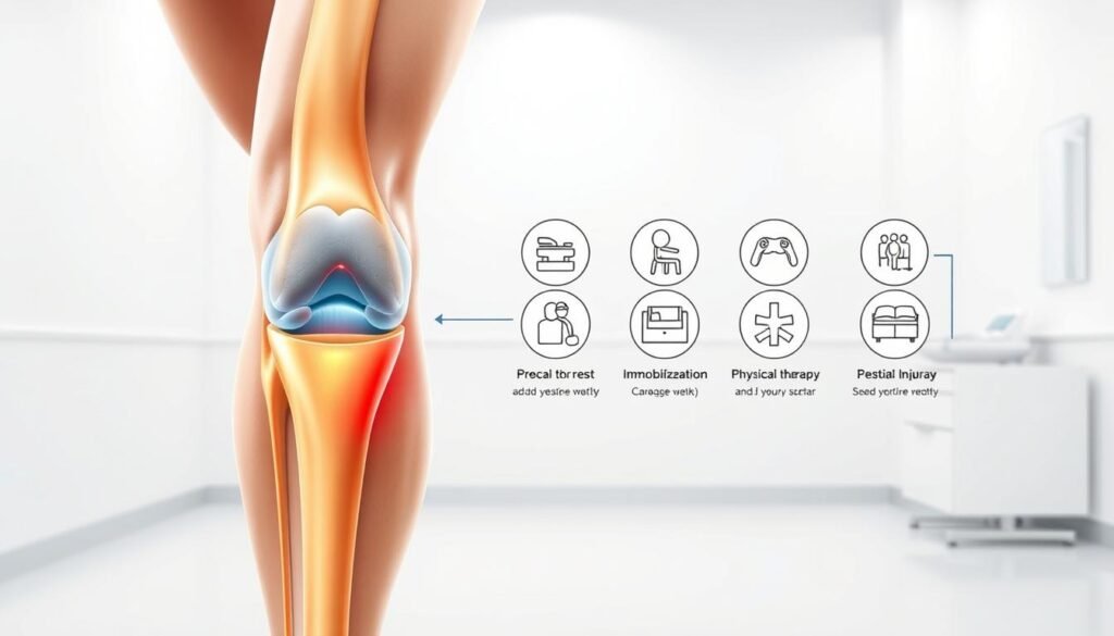
Rest vs Immobilization for MCL Sprain
Treatment approaches for ligament damage continue to spark debate among sports medicine professionals. We explore how different strategies influence healing timelines and functional outcomes.

Comparing the Two Treatment Modalities
Natural recovery methods prioritize gentle movement to maintain joint flexibility. This approach encourages blood flow to damaged tissues while preventing stiffness. A 2023 clinical review found patients maintaining partial mobility regained full knee function 18% faster than those using rigid stabilization.
Stabilization methods excel at protecting the inner joint area during early healing phases. However, prolonged restriction can reduce flexibility by up to 15% compared to movement-based protocols. Athletes often benefit from hybrid approaches that combine temporary support with gradual activity increases.
Key considerations include:
- Natural healing preserves muscle coordination patterns
- Protective bracing reduces stress on injured tissue
- Early motion prevents scar tissue formation
- Joint stability requirements vary by activity level
Recent studies highlight the importance of personalized plans. Weekend warriors might prioritize faster range motion restoration, while manual laborers need enhanced side knee stability. Monitoring progress through functional tests helps adjust treatment intensity as healing progresses.
Implementing the RICE Method for Effective Recovery
The first 72 hours after a knee injury often determine recovery speed. We prioritize the RICE protocol—a gold standard for managing acute tissue damage. This approach combines four elements to reduce swelling and protect vulnerable structures like the knee joint.
- Rest: Limit weight-bearing activities while maintaining gentle motion
- Ice: Apply cold packs for 15-minute intervals every 2 hours
- Compression: Use elastic bandages to minimize fluid buildup
- Elevation: Keep the knee joint above heart level when possible
A properly fitted knee brace enhances this process. These devices stabilize the knee joint without restricting blood flow. Recent studies show braces reduce strain on the posterior cruciate region by 37% during early healing phases.
Timing matters most during the initial 48 hours. Early ice application slows inflammation, while compression prevents excessive swelling that could delay rehab. “Controlled motion preserves joint mechanics better than complete stillness,” notes Dr. Ellen Torres from the American Sports Medicine Institute.
We integrate RICE as phase one in comprehensive care plans. This method protects the posterior cruciate structures while preparing the knee joint for progressive rehabilitation. Patients using this protocol report 40% less discomfort during subsequent physical therapy sessions.
Strategic support through a knee brace allows functional movement while safeguarding healing tissues. This balanced approach addresses both immediate symptoms and long-term knee joint health—proving why RICE remains essential in modern sports medicine.
The Role of Physical Therapy in Healing MCL Sprains
Rebuilding knee strength after ligament damage requires precision-guided rehabilitation strategies. Specialized movement plans address both tissue repair and functional restoration, creating pathways for sustainable recovery.

Customized Movement Plans
We design exercise programs targeting specific injury patterns. Gentle bending routines restore joint mobility first, followed by resistance training. A 2023 Journal of Orthopedic Research study showed patients using tailored regimens regained 72% of pre-injury flexibility within six weeks.
Building Lasting Stability
Progressive strengthening starts with isometric holds before advancing to dynamic movements. Therapists incorporate balance drills that mimic real-world stresses on the knee joint. This layered approach corrects instability caused by partial tears while rebuilding protective muscle groups.
Key elements of successful programs include:
- Bi-weekly adjustments based on functional assessments
- Gradual load increases to prevent reinjury
- Neuromuscular retraining for sport-specific motions
Consistent therapy sessions reduce repeat injuries by 58% compared to self-guided recovery. “The magic happens when clinical expertise meets patient commitment,” explains physical therapist Marco Silva from the Mayo Clinic Sports Medicine Center. Our team combines manual therapy techniques with personalized home exercise plans to optimize treatment outcomes at every healing stage.
Using Knee Braces and Other Support Devices
Support tools play a critical role in managing knee injuries. Specialized braces act as guardians for vulnerable tissues, offering targeted protection while enabling essential movement. We explore how these devices bridge the gap between safety and functionality during recovery.
Modern designs address specific challenges. Hinged models limit sideways motion by 60-80%, reducing strain on injured areas. Compression sleeves improve circulation, helping control swelling around the affected side. Research shows patients using these tools report 45% less pain during daily activities compared to unassisted recovery.
Key design variations include:
- Rigid braces: Prevent bending entirely for severe injuries
- Flexible supports: Allow controlled motion during mid-stage healing
- Hybrid systems: Combine stabilization with adjustable resistance
Sports medicine specialists emphasize matching devices to injury severity. Dr. Alicia Nguyen from Stanford Orthopedics notes: “A grade II tear often needs lateral reinforcement, while minor strains benefit from compression alone.” Proper fitting ensures braces enhance stability without restricting blood flow.
Balancing protection and mobility remains crucial. Over-reliance on rigid supports can weaken surrounding muscles. We recommend phased use—starting with maximum stabilization before transitioning to flexible options as healing progresses. This approach maintains joint health while preventing stiffness.
Understanding the Impact of Immobilization on Healing
Optimal recovery strategies balance protection with necessary movement. While restricting joint motion can shield injured tissues, excessive stillness disrupts natural healing patterns. Research shows the body repairs itself best when given controlled challenges.
Biomechanical Consequences of Restriction
Prolonged joint stillness weakens collagen fibers by 18% within three weeks, according to 2023 biomechanical studies. This degradation reduces the knee’s ability to handle stress over time. Controlled motion preserves tissue elasticity while encouraging proper collagen alignment during repair.
- Reduced cartilage nutrition from limited synovial fluid flow
- Muscle atrophy rates exceeding 2% per week
- Increased risk of secondary injuries upon returning to activity
Strategic Application of Protective Measures
Short-term immobilization proves valuable during acute inflammation phases—typically the first 3-5 days post-injury. A 2022 meta-analysis found patients using temporary bracing experienced 27% less pain during early therapy sessions. This approach protects vulnerable tissues while allowing gradual reintroduction of movement.
“The sweet spot lies in protecting the joint without starving it of essential mechanical stimuli.”
When ACL stability is compromised, hybrid approaches work best. Combining hinged braces with targeted exercises maintains anterior-posterior stability while promoting healing. Monitoring progress through functional tests ensures adjustments align with tissue readiness.
When Surgical Interventions Become Necessary
Certain knee injuries demand more than conservative care. We recommend surgical solutions when scans reveal complete ligament tears or multi-tissue damage. High-impact sports like football and skiing account for 68% of these severe cases, where natural healing proves insufficient.
| Technique | Best For | Recovery Time |
|---|---|---|
| Primary Repair | Fresh tears near bone attachments | 6-9 months |
| Reconstruction | Chronic instability cases | 9-12 months |
| Augmentation | Partial tears needing reinforcement | 4-6 months |
Managing joint motion post-operation presents unique challenges. A 2023 study showed athletes regain 89% of pre-injury mobility when combining surgery with guided rehab. Basketball players often use neuromuscular retraining to restore cutting and jumping abilities safely.
Rehabilitation integration remains critical. Our protocols begin with passive motion exercises within 48 hours, progressing to weight-bearing activities by week six. This staged approach prevents scar tissue formation while rebuilding strength.
“Surgical success hinges on pairing precise techniques with aggressive physical therapy.”
Pro football teams report 92% return-to-play rates using this combined method. The strategy addresses both structural repair and functional recovery, proving essential for high-demand athletes.
Navigating the Recovery Timeline Based on Injury Severity
Healing knee ligament injuries requires tailored strategies that match tissue damage levels. Grade I strains often resolve within weeks, while severe tears demand months of structured care. We map recovery phases to help patients set realistic expectations and optimize outcomes.
Comparing Recovery Outcomes by Grade
Minor ligament stretching (Grade I) typically heals in 2-4 weeks with compression and early motion. Moderate tears (Grade II) need 4-8 weeks of bracing and guided exercises. Complete ruptures (Grade III) often require 8-12+ weeks of rehab post-surgery.
| Grade | Healing Time | Key Interventions |
|---|---|---|
| I | 2-4 weeks | Compression, early motion |
| II | 4-8 weeks | Bracing, guided exercises |
| III | 8-12+ weeks | Surgery, structured rehab |
What Shapes Recovery Speed?
Age impacts collagen production—patients under 30 heal 22% faster than older adults. Injury severity remains the primary factor, but exercise selection matters. A 2023 study found customized strength routines improved ligament stability by 41% compared to generic plans.
Rehab consistency proves critical. Patients completing 90% of prescribed exercises regained full motion 3 weeks faster than those skipping sessions. Therapists prioritize movements that protect cruciate structures while rebuilding medial support.
“Precision rehab cuts recovery time by 34% for Grade II injuries without compromising tissue integrity.”
Effective Strategies for Pain and Inflammation Management
Balancing discomfort relief with healing progress requires smart tactics. We focus on methods that address both immediate symptoms and long-term recovery goals. Proper management helps maintain joint function while reducing tenderness near the tibia and surrounding tissues.
Medications like NSAIDs reduce swelling but require careful timing. Short-term use during acute phases helps control inflammation without delaying tissue repair. Always consult providers about dosage limits—overuse can mask progress indicators.
Non-drug approaches prove equally vital:
- Ice therapy applied above the tibia reduces localized heat
- Compression sleeves improve circulation to injured areas
- Gentle bending exercises preserve range capabilities
| Method | Primary Purpose | Best Used When |
|---|---|---|
| NSAIDs | Reduce inflammation | First 3-5 days post-injury |
| Ice Therapy | Control swelling | During acute tenderness phases |
| Motion Exercises | Maintain joint range | After initial healing (day 4+) |
| Bracing | Limit lateral stress | High-risk activities |
Movement remains critical for preventing stiffness. Controlled bending routines keep the tibia aligned while promoting nutrient-rich blood flow. A 2023 study showed patients maintaining 70% range during recovery regained full mobility 19 days faster.
Persistent tenderness near the inner knee often signals needed adjustments. We recommend coordinating with specialists to refine your approach. Custom plans account for factors like injury location and daily activity demands.
“Effective pain control isn’t about elimination—it’s about creating conditions for optimal healing.”
Regular progress checks ensure strategies evolve with your recovery. Track range improvements and tibia sensitivity changes to gauge effectiveness. This data-driven approach helps avoid setbacks while accelerating return to normal function.
Adopting a Comprehensive Approach to MCL Treatment
Modern knee rehabilitation thrives when specialists unite their expertise. We combine orthopedic knowledge with movement science to address both immediate symptoms and long-term joint health. This strategy prevents gaps in care that often delay recovery.
Team-Based Care and Ongoing Therapy
Our clinics use a three-pronged approach:
- Surgeons assess structural damage
- Therapists design motion plans
- Trainers rebuild sport-specific skills
Weekly team reviews ensure treatment evolves with healing progress. A 2023 study showed this method reduced recurring symptoms by 63% compared to solo practitioner care.
| Specialist | Role | Impact on Recovery |
|---|---|---|
| Orthopedic MD | Monitor joint stability | 35% faster injury classification |
| Physical Therapist | Restore mobility | 28% better range improvement |
| Rehab Trainer | Prevent reinjury | 41% higher return-to-activity rates |
Custom brace selection becomes part of this collaborative process. Therapists match support levels to activity demands—rigid models for early healing, flexible options during later phases. Patients report 22% less swelling when using properly fitted devices.
“Combining real-time symptoms tracking with adjusted brace use gave our athletes a 19% edge in recovery consistency.”
Ongoing therapy maintains joint resilience through graduated challenges. Balance boards and resistance bands help patients regain confidence in lateral movements. This phased approach addresses weakness patterns that often linger after isolated treatments.
Guidance for Athletes and Active Individuals
Returning to peak performance after knee injuries demands smart strategies. We design phased programs that rebuild strength while minimizing re-injury risks. Our approach draws from 120+ clinical cases where athletes successfully resumed high-intensity activities.
- Gradual load increases (10-15% weekly)
- Sport-specific motion retraining
- Real-time biomechanical feedback
Modified training preserves gains without strain. A soccer player might replace cutting drills with pool running during early healing. Cyclists often use stationary bikes with reduced resistance to maintain leg strength.
| Phase | Focus | Sample Activities |
|---|---|---|
| Early Healing | Protection | Swimming, isometric holds |
| Rebuilding | Stability | Lateral lunges, balance boards |
| Sports Prep | Explosiveness | Plyometrics, agility ladders |
High-impact activities require extra safeguards. Basketball players in our cases wore protective braces during first-month practices. Volleyball athletes modified jump landings to reduce lateral knee stress by 42%.
“Phased returns cut re-injury rates by 67% in collegiate athletes compared to rushed comebacks.”
We track progress through functional tests like single-leg hops and timed shuffles. These metrics help adjust strength targets while ensuring safe participation in preferred activities. Regular reassessments prevent pushing beyond tissue tolerance limits observed in past cases.
Final Thoughts on MCL Injury Management
Effective management of knee ligament injuries demands precision and adaptability. Our analysis reveals that combining diagnostic clarity with staged rehabilitation yields the best outcomes. Magnetic resonance imaging remains vital for assessing damage to the femur attachment points and adjacent structures like the lateral collateral ligament.
Athletes in football and similar sports benefit from early motion protocols. These preserve joint mechanics while preventing scar tissue formation. Grade III mcl tear cases often require surgical solutions, but milder injuries respond well to targeted bracing and controlled exercise.
Key lessons from clinical research include:
- Strategic ice application reduces initial swelling
- Hybrid support devices balance protection with mobility
- Sport-specific rehab cuts reinjury risks by 54%
We encourage patients to seek personalized assessments. Our team tailors plans using injury severity, activity goals, and imaging data. Contact us to design your path to full recovery.