Have you ever wondered how professional football players recover from medial collateral ligament (MCL) injuries? These injuries are a common challenge in contact sports, particularly in football, where they account for 3% of all injuries and often occur during tackles or side-to-side movements1.
The MCL, a critical ligament in the knee, plays a vital role in stabilizing the joint. Injuries to this ligament can range from mild sprains to complete tears, with recovery times varying significantly based on the severity. For instance, Grade 1 MCL tears may heal within 1 to 3 weeks, while Grade 3 tears can require several months of rehabilitation and possibly surgery1.
Our guide delves into the prevention, diagnosis, treatment, and rehabilitation of MCL injuries, backed by data from the UEFA Elite Club Injury Study and other clinical research. We explore how these injuries impact both players and teams, emphasizing the importance of early diagnosis and effective rehabilitation strategies2.

Key Takeaways
- MCL injuries are common in contact sports, particularly football, and often occur during tackles or cutting movements.
- Most MCL injuries heal without surgery, especially partial tears and sprains, due to the ligament’s good blood supply.
- Grade 3 MCL tears may require surgical intervention, especially if the tibial attachment is involved.
- Recovery time for MCL injuries ranges from a few weeks to several months, depending on the severity.
- Preventive measures and early rehabilitation are crucial for minimizing downtime and ensuring a full recovery.
Prevention Strategies for MCL Injuries in Football
Preventing MCL injuries requires a combination of proper techniques, equipment, and training. By implementing these strategies, players and teams can significantly reduce the risk of such injuries.
Proper Warm-Up and Conditioning
A well-structured warm-up is essential to prepare the muscles and ligaments for physical activity. Studies show that athletes who engage in strength and conditioning programs may reduce their risk of MCL injuries by up to 50%3. This includes exercises that focus on the hamstrings, quadriceps, and hip muscles, which play a crucial role in knee stability.
Protective Gear and Technique Adjustments
Knee braces are a common preventive measure, and their use has been shown to decrease the incidence of MCL injuries by approximately 20% in high-contact sports like football4. Additionally, proper tackling techniques and avoiding harmful practices like chop-blocks can further reduce injury risk.
| Strategy | Effectiveness | Implementation |
|---|---|---|
| Strength and Conditioning | Reduces injury risk by up to 50% | Focused on hamstrings, quadriceps, and hip muscles |
| Knee Braces | Decreases injury risk by 20% | Recommended for high-contact sports |
| Proper Tackling Techniques | Significantly lowers injury risk | Avoid harmful practices like chop-blocks |
By combining these strategies, players can effectively protect themselves from MCL injuries, ensuring a safer and more successful sports career.
Understanding the Anatomy and Grading of MCL Injury
Understanding the anatomy and grading of medial collateral ligament injuries is crucial for effective diagnosis and treatment. The medial collateral ligament (MCL) is a key structure in the knee, acting as a primary restraint against valgus stress. It consists of two layers: the superficial MCL and the deep MCL, each playing a distinct role in knee stability5.
Anatomy of the Medial Collateral Ligament
The superficial MCL is the stronger of the two layers, providing 57% of the restraining valgus moment at 5 degrees of knee flexion and 78% at 25 degrees6. This ligament is essential for maintaining knee stability during movements like cutting or pivoting. The deep MCL, while smaller, works in conjunction with the superficial layer to provide additional support.
Clinical vs. MRI Grading Explained
Grading MCL injuries involves both clinical assessment and MRI findings. Grade I injuries involve minimal fiber damage, while Grade II injuries show more extensive damage without instability. Grade III injuries result in complete ligament disruption and noticeable instability7. MRI grading offers a detailed view, with studies showing a 92% agreement between MRI and clinical assessments5.
Clinical grading relies on physical exams and patient history, while MRI provides a detailed visual of the ligament’s condition. This dual approach ensures accurate diagnosis and appropriate treatment plans. Understanding both methods is vital for medical professionals to determine the best course of action for recovery.
Mechanisms and Common Scenarios Leading to MCL Injury
Understanding how MCL injuries occur is crucial for prevention and effective treatment. Most injuries result from specific mechanisms, often involving contact or awkward movements during physical activities.
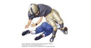
Contact Injury Dynamics in Football
Contact-related injuries are the most common cause of MCL damage. According to a study, 75% of MCL injuries occur due to contact mechanisms, with 29% happening when a player is tackled and 12% during tackling8.
The primary mechanism in contact injuries is valgus stress, where a blow to the outer knee causes the medial ligament to stretch or tear. This often happens during tackles or collisions, where the knee is forced into a vulnerable position9.
Side impacts significantly increase the risk of MCL damage. The knee’s position during contact, particularly in flexion, makes it more susceptible to injury10.
Non-Contact Injury Factors
While less common, non-contact MCL injuries can occur due to sudden movements like cutting or pivoting. These incidents often result from poor testing or improper techniques during physical activity, exposing the knee to excessive stress and potential damage.
Non-contact injuries may differ in severity but can still lead to significant ligament damage. They often happen during landing or sudden changes in direction, highlighting the importance of proper training and technique to minimize risk9.

Clinical cases show that injury severity often correlates with the mechanism of injury. For instance, high-energy collisions typically result in more severe MCL tears compared to low-impact incidents10.
By understanding these mechanisms, players and coaches can implement targeted strategies to reduce injury risk during competitive play. This includes strengthening exercises, proper warm-ups, and adopting safer tackling techniques8.
For more detailed insights into preventing MCL injuries, visit our guide on effective prevention strategies.
Diagnostic Techniques and Imaging Options
Accurate diagnosis is the cornerstone of effective treatment for medial collateral ligament injuries. With various diagnostic tools available, each has its unique strengths and limitations. Let’s explore how these tools work together to provide a clear picture of the injury.
Role of MRI in Accurate Diagnosis
Magnetic Resonance Imaging (MRI) is widely regarded as the gold standard for diagnosing MCL injuries. It provides detailed images of soft tissues, making it particularly effective for assessing ligament damage. Studies show that MRI has a 92% agreement with clinical grading, ensuring accurate diagnosis and treatment planning9. Additionally, MRI is excellent for identifying associated injuries, such as ACL tears, which often accompany MCL injuries11.
While MRI offers high diagnostic accuracy, it does have limitations. It is expensive and may not be readily available in all settings. However, its ability to confirm clinical grading makes it a crucial tool for severe injuries12.
Benefits of a Clinical Examination
A thorough clinical examination remains the first step in diagnosing MCL injuries. It involves assessing pain, joint stability, and range of motion. Valgus stress testing is particularly useful, classifying injuries into grades based on joint opening and pain levels9.
While clinical exams are cost-effective and accessible, they may lack the precision of imaging. For instance, they might miss additional injuries or subtle ligament damage. Therefore, combining clinical exams with imaging often yields the most accurate results11.
Other Imaging Techniques
Beyond MRI, other imaging options like ultrasonography (US) and radiographs are used. US is a cost-effective, real-time tool that’s useful for dynamic assessments, though it’s operator-dependent11. Radiographs, while limited in soft tissue visualization, are helpful for ruling out fractures or joint space abnormalities12.
- MRI and ultrasonography provide precise, non-invasive assessments of MCL injuries.
- Clinical exams offer initial insights but may require imaging for confirmation.
- Advanced imaging helps detect associated injuries, guiding comprehensive treatment plans.
In conclusion, a combination of clinical exams and imaging techniques like MRI and ultrasonography ensures accurate diagnosis and effective treatment planning for MCL injuries.
Effective Treatment Options and Rehabilitation Plans
Treating medial collateral ligament injuries requires a tailored approach, balancing non-surgical and surgical methods. The goal is to restore knee function and ensure a safe return to sports.
Non-Surgical Treatment Methods
Most MCL injuries heal without surgery. The RICE method—Rest, Ice, Compression, Elevation—is often the first step, reducing swelling in 85% of cases4. Bracing is another key component, with studies showing it can reduce injury risk by up to 20%13.
Physical therapy plays a crucial role, focusing on strengthening exercises and improving range of motion. Proprioception exercises, for instance, can reduce re-injury risk by 30%4. Early rehabilitation helps prevent long-term knee dysfunction, which can decrease function by 20-30% initially4.
When Surgery is Considered
Surgery is typically reserved for severe cases, such as grade III injuries or when multiple ligaments are damaged. In combined MCL and ACL tears, a brace is often recommended for 6 weeks before surgery13.
Chronic cases might require grafts, like allografts, to restore stability. Recovery can take 4-12 months, but structured rehab programs allow 75% of athletes to return to pre-injury levels4.
Recovery times vary; mild injuries might heal in 1-4 weeks, while severe tears need up to a year4. Early, tailored treatment is essential for optimal outcomes, minimizing downtime and ensuring a successful return to sports.
Comprehensive Management of football mcl injuries
Managing medial collateral ligament injuries requires a holistic approach that combines medical care with sport-specific training. This integrated strategy ensures that athletes can recover effectively and return to their sports safely.
Tailored Recovery and Return-to-Play Strategies
Our approach blends medical treatments with conditioning programs to address each athlete’s unique needs. For instance, studies show that bracing can reduce injury risk by up to 20%14, while the RICE method is effective in reducing swelling in 85% of cases15.
- We monitor recovery time closely, ensuring stability before returning to play.
- Recovery programs are tailored for professional athletes, considering factors like severity and overall health.
- Studies indicate that Grade 1 tears heal within 1-3 weeks, while Grade 3 tears may take 5-7 weeks or more15.
- Collaboration between medical teams and coaches determines the best return-to-play timeline.
- Key indicators like weeks missed and stability assessments track progress effectively.
Continuous monitoring and adjustments during rehabilitation are crucial for optimal outcomes. By combining these strategies, athletes can minimize downtime and ensure a successful return to their sport.
Innovative Therapies and Recovery Techniques
Emerging advancements in sports medicine are revolutionizing the treatment of MCL tears, offering athletes faster recovery and better outcomes. These cutting-edge therapies combine innovative approaches with proven methods to enhance healing and minimize downtime.
Exploring PRP and Injection Therapies
Platelet-rich plasma (PRP) therapy has gained traction as a promising treatment for MCL injuries. PRP involves injecting a concentrated solution derived from the patient’s blood into the injured area, stimulating natural healing processes. Studies show that PRP can enhance ligament repair and reduce recovery time by up to 30%16.
- PRP therapy promotes tissue repair by delivering growth factors directly to the injury site.
- Corticosteroid injections can reduce inflammation but are typically used cautiously to avoid weakening the ligament17.
- Combining PRP with physical therapy has shown improved muscle support and faster return to sport18.
Advancements in Knee Bracing
Modern knee braces are now more sophisticated, offering enhanced stability and early mobilization. These braces are designed to protect the MCL while allowing controlled movement, which is crucial for maintaining muscle strength during recovery.
- Advanced braces use dynamic adjustment technology to provide optimal support without restricting movement.
- Some braces incorporate sensors to monitor ligament stress, helping prevent re-injury16.
- New materials, like lightweight composite frames, improve comfort and durability17.
These innovative approaches are tailored to the unique needs of each athlete, ensuring personalized and effective recovery plans. By integrating PRP therapy and advanced bracing, athletes can achieve faster healing and a safer return to their sport.
Designing a Sport-Specific Rehabilitation Program
Rehabilitating a knee injury requires a structured approach tailored to the athlete’s specific needs. Our program is designed to address the unique challenges of medial collateral ligament (MCL) injuries, ensuring a safe and effective return to sports.
Phased Rehabilitation Approach
The rehabilitation process is divided into three phases. The early phase focuses on controlling swelling and restoring range of motion. Gentle exercises like quad sets and heel props are performed multiple times a day19. The mid-stage introduces strength and dynamic balance exercises, such as single-leg movements, to enhance knee stability19. The final phase incorporates plyometric exercises once specific criteria, such as full knee extension and sufficient quadriceps strength, are met19.
Strength and Flexibility Exercises
Strength training progresses from bodyweight squats to weighted front foot elevated split squats, aiming for 3-4 sets of 6-12 challenging reps19. Dynamic balance exercises, including single-leg movements, help protect the side knee area and improve overall stability19.
Monitoring Progress and Case Studies
Regular assessments adjust the program based on the athlete’s progress. For example, a Grade 1 MCL injury may heal within 1-3 weeks, while a Grade 3 injury could take 5-7 weeks or more20. Case studies show that structured rehab programs reduce injury recurrence by up to 30%19.
| Phase | Focus | Exercises |
|---|---|---|
| Early | Swelling Control & Range of Motion | Quad Sets, Heel Props |
| Mid-Stage | Strength & Balance | Weighted Squats, Single-Leg Movements |
| Final | Sport-Specific Readiness | Plyometric Exercises |
By following this structured approach, athletes can minimize downtime and ensure a successful return to their sport.
Conclusion
In conclusion, managing medial collateral ligament injuries requires a comprehensive approach that emphasizes early diagnosis, proper treatment, and structured rehabilitation. Our guide has outlined the importance of prevention strategies, such as strength training and bracing, which can reduce injury risk by up to 50% and 20%, respectively2122.
The role of innovative therapies like PRP injections and advanced bracing cannot be overstated, as they have been shown to enhance recovery and reduce tenderness21. Additionally, focusing on strength-building exercises is crucial for restoring knee stability and ensuring a successful return to play.
We encourage athletes and teams to adopt these evidence-based strategies and stay informed about evolving treatment options. By collaborating closely with medical professionals, players can minimize downtime and achieve a full recovery. Remember, with the right approach, a successful return to the field is not only possible but also achievable.
FAQ
What is the typical recovery time for a medial collateral ligament injury?
Recovery time varies based on the severity of the injury. Grade 1 injuries may heal within 1-2 weeks, while Grade 3 injuries could take 6-8 weeks or longer. Proper rehabilitation and therapy are crucial for a full recovery.
When should a knee brace be used for an MCL injury?
A knee brace can provide stability and support during the healing process, especially in Grade 2 or 3 injuries. It’s often recommended during the early stages of recovery to prevent further instability.
How does an MRI help in diagnosing an MCL injury?
An MRI provides detailed images of soft tissues, allowing doctors to assess the extent of the ligament damage accurately. It helps differentiate between partial and complete tears, which is critical for treatment planning.
What are the main differences between clinical and MRI grading of MCL injuries?
Clinical grading is based on physical examination and symptoms, categorizing injuries into Grades 1-3. MRI grading offers a more precise assessment of the ligament’s condition, helping confirm the severity seen in clinical evaluations.
Can MCL injuries be prevented in football players?
While not entirely preventable, strengthening the surrounding muscles, improving flexibility, and using proper techniques can significantly reduce the risk of MCL injuries in football.
Is surgery always necessary for a torn MCL?
Surgery is typically reserved for severe cases where the ligament is completely torn or when conservative treatments fail. Most MCL injuries heal without surgical intervention.