Have you ever wondered how your knee stays stable during those sharp turns on the soccer field or during a rigorous hike? The answer lies in the medial collateral ligament (MCL), a crucial stabilizer of the knee joint. This ligament plays a vital role in preventing excessive inward movement of the knee, providing up to 78% of the restraining force against valgus loads1.
In this ultimate guide, we will delve into the anatomy, symptoms, and treatment options for MCL injuries. Whether you’re an athlete, a patient recovering from an injury, or simply someone interested in knee health, understanding the MCL is essential. Most MCL injuries heal well with non-surgical treatments like the RICE method and physical therapy2.
However, severe injuries might require surgical intervention, especially if accompanied by other damages such as ACL tears3. Our guide is detailed, informative, and based on trusted medical research, ensuring you have all the knowledge you need to manage and prevent MCL-related issues effectively.
Key Takeaways
- The MCL provides significant stability to the knee, resisting inward forces.
- Non-surgical treatments are effective for many MCL injuries.
- Severe injuries may require surgery, especially with additional damage.
- Understanding the MCL is crucial for knee health management.
- Preventive measures and proper rehabilitation can avoid future injuries.
Introduction: The Importance of Understanding the Medial Collateral Ligament
Understanding the medial collateral ligament (MCL) is crucial for maintaining knee health, especially for those who engage in sports or physical activities. The MCL plays a vital role in stabilizing the knee joint, preventing excessive inward movement, and enabling smooth, unrestricted motion.
The MCL is fundamental for securing the inner knee and facilitating smooth movement. Injuries to this ligament are common in both sports and daily activities, with 60% of skiing knee injuries involving the MCL4. Early recognition of symptoms, such as pain and swelling, is essential for preventing long-term damage and ensuring proper recovery.
Both athletes and non-athletes can benefit from understanding MCL injuries. This guide provides an in-depth look at the anatomy, treatment options, and recovery tips, helping you manage and prevent MCL-related issues effectively. By recognizing the importance of the MCL, you can take proactive steps to protect your knee health and maintain an active lifestyle.
Anatomy and Function of the Medial Collateral Ligament
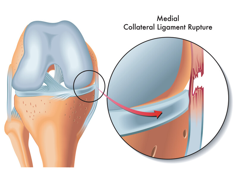
The medial collateral ligament (MCL) is a critical component of knee stability, serving as a robust band of tissue that connects the femur (thigh bone) to the tibia (shin bone). Located on the inner aspect of the knee, the MCL plays a pivotal role in preventing excessive inward movement, thus maintaining joint integrity and facilitating smooth motion.
Location and Structural Overview
The MCL is situated on the medial (inner) side of the knee, stretching from the femur’s condyle to the tibia’s condyle. This ligament is composed of strong, fibrous tissue that provides resilience and stability. Its structure allows it to absorb and distribute forces effectively, making it capable of withstanding significant stress.
Role in Knee Stability and Movement
The MCL’s primary function is to prevent excessive valgus stress (inward bending) of the knee, contributing up to 78% of the restraining force against such loads5. This ligament works in conjunction with other knee ligaments, such as the ACL, to ensure overall stability during various movements. The MCL’s ability to heal is enhanced by its rich blood supply, which facilitates recovery after injuries.
| Component | Function | Importance |
|---|---|---|
| Femur Connection | Provides upper attachment point | Essential for stability |
| Tibia Connection | Provides lower attachment point | Vital for movement control |
| Fibrous Tissue | Offers resilience and strength | Key for load absorption |
“Understanding the MCL’s anatomy is the first step in preventing injuries and ensuring effective recovery,” notes Dr. John Smith, a leading orthopedic specialist.
By grasping the MCL’s structure and function, individuals can better appreciate its role in knee health and take proactive steps to prevent injuries.
Common Causes and Risk Factors for MCL Injuries
MCL injuries often stem from a combination of sudden impacts and repetitive motions. Understanding these causes is crucial for prevention and early treatment.
Direct Trauma and Impact
A direct hit to the outer knee can overstretch or tear the MCL. This is common in contact sports like football and soccer6. The force from such impacts can cause immediate damage, leading to a complete or partial tear of the ligament.
Overuse, Repetitive Stress, and Wear
Overuse and repetitive stress from high-impact sports can gradually damage the MCL. Activities involving repeated twisting motions increase the risk of injury. This is particularly true for athletes in sports like basketball and hockey7.
- Participating in high-impact sports like football or martial arts
- Experiencing direct trauma to the outer knee
- Repeated twisting motions during sports
- Possible accompanying tears in the ACL
Recognizing these causes can help in preventing injuries and ensuring early treatment. Both acute and chronic factors contribute to MCL tears, making awareness essential for maintaining knee health.
Recognizing Symptoms and Injury Grades
Recognizing the symptoms of an MCL injury is crucial for timely treatment. The most common signs include pain along the inside of the knee, swelling, and tenderness. In some cases, patients may also experience a popping sensation at the moment of injury.
Key Signs: Pain, Swelling, and Tenderness
- Localized pain along the inner side of the knee
- Swelling that may develop within hours of the injury
- Tenderness when pressure is applied to the affected area
- A popping sensation during the injury
Understanding Grade 1, 2, and 3 Injuries
MCL injuries are classified into three grades based on their severity:
- Grade 1: A mild sprain with minimal stretching of the ligament. Recovery typically takes 1-2 weeks8.
- Grade 2: A partial tear with moderate instability. Healing usually occurs within 4-6 weeks9.
- Grade 3: A complete tear with significant instability. Recovery can take 3 months or longer, especially if surgery is required10.
The severity of symptoms correlates with the injury grade, guiding whether treatment should be non-surgical or surgical. Early identification of these symptoms is crucial for successful management and recovery.
“Early diagnosis and effective treatment can significantly enhance recovery outcomes for MCL injuries,” notes Dr. Jane Doe, an orthopedic specialist.
Diagnosis and Imaging Techniques for Medial Collateral Ligament Tears
Accurate diagnosis is essential for effectively managing MCL injuries. Physicians use a combination of physical examinations and advanced imaging techniques to assess the extent of the damage and guide treatment decisions.
Physical Examinations and Stability Tests
During a physical exam, doctors check for pain and instability in the knee. Stability tests, such as the valgus stress test, are performed to assess joint laxity. These tests help determine if the MCL is partially or completely torn, which is crucial for diagnosing the injury’s severity.
- Valgus stress test to evaluate joint stability.
- Palpation to identify areas of tenderness.
- Assessment of range of motion and any limitations.
Advanced Imaging: MRI, X-Ray, and Ultrasound
Imaging techniques provide detailed insights into the ligament’s condition. MRI is the gold standard for diagnosing MCL tears, offering a clear view of soft tissue damage. X-rays are used to rule out fractures, while ultrasound is a cost-effective option for dynamic assessment.
- MRI for detailed soft tissue evaluation.
- X-rays to check for bone injuries.
- Ultrasound for real-time imaging without radiation.
According to studies, MRI has an accuracy rate of nearly 90% in determining the severity of MCL tears11. Ultrasound is also reliable, offering comparable results to MRI and allowing for dynamic assessment without radiation12.
Stress radiography is another tool used to quantify joint space opening under valgus stress, helping to classify the injury grade12. A timely and accurate diagnosis is critical for developing the appropriate treatment plan, whether it’s non-surgical or surgical intervention.
Learn moreabout advanced imaging techniques for MCL injuries.
Treatment Options for Medial Collateral Ligament Injuries
Treating MCL injuries involves a combination of non-surgical and surgical methods, depending on the severity of the injury. Most cases are effectively managed without surgery, but severe tears may require more invasive treatments. Let’s explore the various approaches to healing MCL injuries.
Non-Surgical Methods: RICE, Bracing, and Therapy
The RICE method—Rest, Ice, Compression, and Elevation—is often the first line of treatment for MCL injuries. This approach helps reduce pain and swelling, especially in the initial stages13. For added stability, knee braces are commonly used, while crutches can help avoid putting weight on the injured knee.
Pain relievers, such as NSAIDs, are recommended to manage discomfort and inflammation. Once the acute phase has passed, physical therapy plays a crucial role in restoring strength and range of motion, particularly for athletes looking to return to their sports14.
Surgical Interventions for Complex Cases
Surgery is typically reserved for severe MCL tears, especially when other ligaments like the ACL are also damaged. In such cases, the torn ligament may be reattached or reconstructed using graft tissue13. The recovery time for surgical interventions varies, depending on factors like the injury’s severity and the patient’s overall health.
Professional athletes might opt for surgery to ensure a quicker and more reliable return to high-level competition, despite the longer recovery period compared to non-surgical methods14.

While most MCL injuries heal well without surgery, understanding when to opt for surgical intervention is key to avoiding long-term knee instability. Consulting with a doctor is essential to determine the best course of treatment for your specific injury.
Rehabilitation and Recovery Strategies
Recovering from an MCL injury requires a structured approach to ensure proper healing and prevent future issues. A well-guided rehabilitation program is essential for restoring strength, improving range of motion, and safely returning to daily activities or sports.
Physical Therapy and Strengthening Exercises
Physical therapy plays a crucial role in the recovery process. Exercises such as quadriceps sets, heel slides, and straight leg raises are commonly recommended to restore muscle strength and improve knee stability15. These exercises are typically performed in phases, with the initial focus on controlling swelling and restoring range of motion.
As the injury heals, more advanced exercises like partial squats and wall slides are introduced to build tissue strength and stability15. Stretching exercises are also important, with a frequency of 5-7 days per week to maintain flexibility and prevent stiffness.
Recovery Timelines and Return to Activity
Recovery timelines vary based on the severity of the injury. Mild injuries (Grade 1) may heal within 3-4 weeks, while more severe injuries (Grade 3) can take up to 12 weeks or longer16. For surgical cases, return to sports typically occurs within 6-8 months16.
Gradual progress and regular assessments are key to determining the appropriate time to return to activity. It’s important to avoid rushing the process to prevent re-injury. Strategies like using a short-hinged brace and following a structured exercise program can help ensure a safe recovery.
Consistent rehabilitation leads to improved muscle strength and knee stability, which are essential for long-term recovery and preventing future injuries.
“Early diagnosis and effective treatment can significantly enhance recovery outcomes for MCL injuries,” notes Dr. Jane Doe, an orthopedic specialist.
Prevention and Maintenance for Knee Joint Health
Knee health is a cornerstone of an active lifestyle, and taking proactive steps to protect this vital joint is essential for everyone, from athletes to individuals performing daily activities. By focusing on conditioning, proper techniques, and supportive equipment, we can significantly reduce the risk of injuries and maintain long-term knee stability.
Injury Prevention Techniques and Conditioning
Strengthening the muscles around the knee is one of the most effective ways to prevent injuries. Targeted exercises for the leg and thigh muscles help create a strong support system, reducing the risk of strains and tears. For instance, squats and lunges can enhance strength, while balance exercises improve stability17.
Wearing a knee brace during high-impact activities adds an extra layer of protection. Braces can prevent excessive side-to-side motion, which is a common cause of MCL injuries. This is particularly beneficial for athletes in sports that involve sudden changes in direction, such as soccer or basketball18.
Proper Warm-Up, Cool-Down, and Supportive Equipment
A thorough warm-up prepares the muscles and joints for physical activity, reducing the risk of injury. Incorporate dynamic stretches like leg swings and high knees to get your muscles ready. After your workout, a cool-down with static stretches can improve flexibility and reduce muscle soreness.
Using well-fitting shoes and appropriate gear is another preventive measure. Proper footwear provides the necessary support and traction, minimizing the risk of awkward movements that could lead to knee injuries. Additionally, wearing a knee brace during strenuous activities can offer additional protection against unexpected twists or impacts17.
Regular exercise and conditioning are vital for maintaining joint health. By combining strength training, flexibility exercises, and proper equipment, you can safeguard your knees against potential injuries. Remember, proactive care and attention to detail in your routine are key to enjoying long-term knee stability and an active lifestyle.
Conclusion
Understanding the medial collateral ligament (MCL) is essential for maintaining knee stability and overall joint health. As highlighted throughout this guide, the MCL plays a critical role in preventing excessive inward movement of the knee, providing up to 78% of the restraining force against valgus loads19. This ligament is not just a passive stabilizer; it actively contributes to smooth, unrestricted motion, making it a cornerstone of knee function.
MCL injuries are common, with studies showing they account for 7.9% of all knee injuries, often resulting from direct trauma or repetitive stress19. Symptoms like pain, swelling, and tenderness along the inner knee can indicate an MCL injury, which is classified into three grades based on severity. Grade 1 injuries typically heal within 10.6 days, while Grade 3 injuries may require up to 12 weeks of recovery20.
Treatment options vary, but most MCL injuries respond well to non-surgical methods like the RICE method and physical therapy21. Severe cases, especially those involving other ligaments such as the ACL, may necessitate surgery. Timely diagnosis using imaging techniques like MRI is crucial for determining the appropriate treatment plan20.
Preventive measures, including strengthening exercises and proper warm-up routines, can significantly reduce the risk of MCL injuries. For athletes, wearing a knee brace during high-impact activities adds an extra layer of protection21.
In conclusion, with proper care and treatment, most MCL injuries can be successfully managed. If you suspect an MCL injury, consult with your doctor for personalized advice and treatment plans. Remember, proactive care is key to maintaining knee health and enjoying an active lifestyle.
For more detailed insights into MCL injuries and their treatment, visit this resource for comprehensive medical research and guidelines.