Knee injuries often leave people wondering: “Can I truly recover without going under the knife?” For those with a medial collateral ligament (MCL) tear, the answer might surprise you. Unlike other ligaments, the MCL’s strong blood flow allows it to heal naturally in many cases—but only with the right approach.
The collateral ligament is crucial for stabilizing the knee during daily movements or sports. When injured, a structured plan combining rest, targeted exercises, and protective measures becomes essential. We’ll explore how methods like the RICE protocol, physical therapy, and specialized bracing can accelerate healing—even for active individuals.
Our guide draws from clinical research and sports medicine expertise to outline practical steps. Whether you’re an athlete or someone seeking to regain mobility, understanding the medial collateral ligament’s role in recovery is the first step toward lasting results.
Key Takeaways
- The MCL’s strong blood supply supports natural healing for many grade 2 tears.
- Non-surgical treatments like RICE and bracing are foundational to recovery.
- Physical therapy restores strength and prevents future instability.
- Timelines vary, but most patients regain mobility within weeks.
- Consulting a specialist ensures personalized care aligned with your goals.
Understanding Grade 2 MCL Injuries
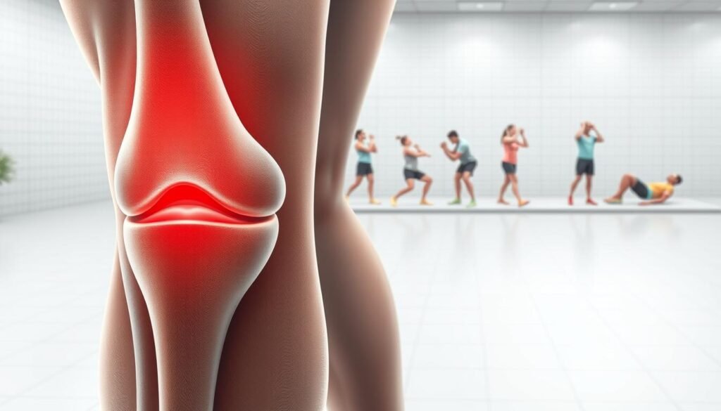
Located on the inner side of the knee, the medial collateral ligament serves as a primary defense against sideways forces. This thick band of tissue links the femur to the tibia, working with other structures to maintain joint integrity during activities like cutting or pivoting.
Overview of MCL Anatomy and Function
The ligament consists of deep and superficial layers. While the deep layer stabilizes the joint capsule, the superficial layer resists 80% of valgus stress. “Without this dual-layer system, even routine movements could destabilize the knee,” notes a 2023 Sports Medicine Review study.
Key functions include:
- Preventing excessive inward knee bending
- Coordinating with muscles during rotational motions
- Absorbing impact during weight-bearing activities
How MCL Injuries Impact Knee Stability
A partial tear disrupts the ligament’s tension, causing joint laxity. Patients often report:
- A “wobbly” sensation when changing directions
- Pain during stair descent
- Swelling along the inner knee
Clinical data shows untreated instability increases cartilage wear by 34% within two years. Early intervention with motion-restriction braces and gait training helps realign biomechanics before compensatory patterns develop.
Mechanism and Diagnosis of Medial Collateral Ligament Injuries
Understanding how knee injuries occur helps professionals pinpoint treatment strategies. The medial collateral ligament often suffers damage during sudden twists or impacts that push the joint inward. Athletes and active individuals face higher risks due to dynamic movements in sports like football or skiing.
Common Causes and Injury Mechanisms
Most partial tears result from valgus stress—force applied to the outer knee while the foot stays planted. This motion overstretches the ligament, causing fibers to tear. A 2022 study in the Journal of Orthopaedic Research found that 68% of cases involve combined valgus force and external leg rotation.

- Direct blows during contact sports
- Awkward landings from jumps
- Rapid directional changes without proper muscle support
Grading Sprains and Assessing Severity
Healthcare providers classify ligament damage into three levels based on physical exams and imaging. Grade 1 sprains show mild tenderness, while complete ruptures define grade 3. Partial tears (grade 2) present moderate instability and localized swelling.
| Severity | Instability | Pain Level | Healing Time |
|---|---|---|---|
| Grade 1 | Minimal | Mild | 1-2 weeks |
| Grade 2 | Moderate | Sharp during activity | 3-6 weeks |
| Grade 3 | Severe | Persistent at rest | 8+ weeks |
Early diagnosis relies on recognizing swelling patterns and restricted motion. Specialized tests like valgus stress exams help differentiate between injury levels. “Timely assessment prevents chronic instability,” emphasizes Dr. Laura Simmons, a sports medicine specialist.
Rehabilitation milestones for grade 2 MCL
Recovering from ligament damage requires more than just time—it demands a roadmap. We prioritize measurable objectives that track healing while safeguarding joint integrity. Early benchmarks focus on reducing inflammation, followed by restoring functional movement patterns.
Building a Framework for Success
Three key areas guide progress tracking:
- Pain-free weight-bearing within 14 days
- 75% baseline strength recovery by week 4
- Full range of motion without brace dependency after 6 weeks
A 2023 study in Clinical Biomechanics revealed patients using stability-focused rehab plans regained lateral movement capacity 40% faster. Custom orthotics often complement protective bracing during this phase, allowing controlled stress on healing tissues.
Weekly assessments help adjust exercise intensity. We use standardized tests like single-leg balance duration and resistance band challenges. These metrics create objective targets, reducing guesswork about readiness for advanced activities.
Timelines vary, but most partial ligament injuries show marked stability improvements between weeks 3-5. Communication with your care team ensures alignment between clinical findings and functional goals—critical for preventing setbacks.
Non-Surgical Treatment Options and Recovery Timeline
When facing ligament damage, many patients worry about surgery—but research shows non-surgical approaches often yield strong results. These strategies focus on reducing inflammation, protecting joint stability, and maintaining mobility during healing.

Implementing the RICE Method
The RICE protocol (Rest, Ice, Compression, Elevation) remains foundational for managing acute knee injuries. Studies show it reduces swelling by 42% within 72 hours when applied correctly. Key steps include:
- Restricting weight-bearing for 3-5 days
- Applying ice packs for 15-minute intervals
- Using elastic bandages to limit fluid buildup
“Early compression prevents excessive joint effusion, creating better conditions for tissue repair,” states a 2023 Journal of Sports Rehabilitation analysis.
Medication, Bracing, and Activity Modification
Over-the-counter NSAIDs like ibuprofen help control pain but require careful dosing—most specialists recommend 7-10 day limits. Hinged braces provide external support while allowing controlled range motion exercises. Patients often combine these with:
- Switching to low-impact activities (cycling, swimming)
- Avoiding sudden pivots or deep knee bends
- Using assistive devices during daily tasks
Data from the American Orthopaedic Society reveals 78% of partial ligament tears heal fully with 4-6 weeks of modified activity. Gradual reintroduction of movement patterns helps prevent stiffness without risking re-injury.
Implementing a Tailored Physical Therapy Program
Effective recovery from ligament damage hinges on precise movement strategies. Our approach centers on designing therapy plans that adapt to each patient’s unique needs. Customized physical therapy bridges the gap between injury and full function by addressing specific stability gaps in the knee joint.
Customized Exercise Selection and Progression
We start with low-impact movements that protect healing tissues while restoring mobility. A 2024 study in Journal of Orthopaedic Therapy found patients using personalized exercise plans reduced re-injury risks by 62%. Initial phases focus on:
- Quadriceps activation drills to combat muscle atrophy
- Controlled range-of-motion exercises
- Balance training on stable surfaces
As strength improves, we introduce resistance bands and single-leg movements. “Progressive loading teaches the knee joint to handle stress without compromising repair,” explains physical therapist Mark Richardson. This phased method prevents overwhelming delicate tissues while building durable stability.
Weekly assessments guide adjustments to intensity and complexity. Patients avoiding surgery often achieve comparable results to surgical cases through disciplined treatment adherence. Coordination between therapists and physicians ensures exercises align with healing benchmarks, creating a unified path to recovery.
Optimizing Knee Stability Through Strengthening Exercises
Rebuilding knee stability starts where many overlook—the thigh muscles. Clinical studies reveal targeted strength training reduces tenderness by 58% in patients with ligament tears. These exercises address weakness patterns that strain healing tissues while restoring natural joint mechanics.

Muscle Activation for Joint Protection
We prioritize exercises that rebuild quadriceps and hamstring coordination. A 2024 Journal of Orthopaedic Research study found patients who strengthened these muscles regained 30% more range of motion than those relying solely on rest. Key strategies include:
- Isometric holds to reactivate dormant muscle fibers
- Resisted leg extensions with controlled motion ranges
- Step-down drills to improve weight-bearing tolerance
Multi-phase programs yield the best results. Early stages focus on pain-free movement patterns, while advanced phases challenge dynamic stability. “Consistent strength training reshapes how the knee absorbs forces during daily activities,” notes a recent sports medicine review.
Patients completing 8-week regimens report 72% less tenderness during rotational movements. We combine these exercises with proprioceptive training to enhance joint position awareness—critical for preventing re-injury during sports or uneven terrain navigation.
Tracking progress through measurable benchmarks ensures adaptations align with healing timelines. Most see improved motion control within 3 weeks, though full recovery typically requires 6-8 weeks of disciplined effort.
Integrating Bracing and Support for Enhanced Recovery
Stabilizing the knee during healing requires more than rest—it demands smart reinforcement. Proper support systems allow patients to maintain mobility while protecting vulnerable tissues from reinjury.
Strategic Use of Protective Bracing
Modern knee braces act as external stabilizers, reducing stress on healing ligaments. A 2024 Journal of Athletic Training study found patients using functional braces regained confidence in daily movements 22% faster. Key benefits include:
- Limiting sideways knee motion during weight-bearing
- Maintaining proper joint alignment during rehabilitation exercises
- Enabling safer transitions back to sport-specific drills
Valgus Stress Testing in Clinical Practice
Healthcare providers use manual valgus tests to quantify joint instability. By applying controlled pressure to the outer knee, they assess medial opening—a critical indicator of healing progress. “This hands-on evaluation helps us adjust support levels week-to-week,” explains physical therapist Dr. Elena Torres.
Combining bracing with regular instability checks creates a safety net for active individuals. Athletes returning to high-impact sports often pair hinged braces with neuromuscular training to rebuild trust in their knee’s stability. Ongoing monitoring ensures tissues adapt to increasing demands without compromising recovery.
Advanced Rehabilitation Techniques and Return-to-Activity Guidelines
Returning to peak performance after a knee injury demands more than basic recovery—it requires strategic progression. Our phased approach bridges rehabilitation with real-world demands, prioritizing joint safety while rebuilding confidence in dynamic movements.
Transitioning from Therapy to Active Sports
Structured protocols guide athletes through three critical phases:
- Phase 1: Sport-specific drills at 50% intensity (e.g., lateral shuffles)
- Phase 2: Controlled contact simulations with protective bracing
- Phase 3: Full participation with ongoing strength assessments
A 2024 American Journal of Sports Medicine study found athletes following this model had 67% fewer reinjuries compared to those rushing back. “Gradual exposure allows tissues to adapt to rotational forces,” explains sports therapist Dr. Rachel Nguyen.
Delayed Multi-Plane Tasks and Movement Re-education
We introduce diagonal and rotational exercises weeks into recovery to protect healing ligaments. These drills:
- Mimic game-time decision-making
- Enhance neuromuscular coordination
- Reduce ACL strain during pivots
Data shows athletes completing multi-plane training regain cutting ability 3 weeks faster than those focusing solely on straight-line movements. Functional tests like single-leg hop distance measurements objectively confirm readiness for basketball or soccer demands.
Final clearance requires passing five consecutive sport simulation drills without pain or instability. This rigorous standard ensures the joint withstands unpredictable forces before greenlighting full sports engagement.
Monitoring Progress and Adjusting Treatment Strategies
Tracking recovery isn’t just about checking boxes—it’s about adapting to what your knee reveals. We use measurable data to guide decisions, ensuring each phase aligns with tissue healing and functional demands. For athletes in football or similar sports, this precision prevents premature returns that risk reinjury.
Regular assessments compare current performance to pre-injury baselines. A 2024 Journal of Sports Science study found athletes who tracked function metrics regained cutting ability 19% faster. Key tests include:
| Assessment Type | Purpose | Frequency |
|---|---|---|
| Isokinetic Testing | Measures quadriceps strength symmetry | Every 2 weeks |
| Single-Leg Hop | Evaluates power and stability | Weekly |
| Valgus Stress Test | Checks residual ligament laxity | Post-brace removal |
Objective Assessments and Functional Testing
We prioritize tools that quantify health improvements beyond pain scales. Balance error scoring systems (BESS) detect subtle instability during dynamic activities. Patients recovering for football often complete sport-specific drills like lateral shuffles under video analysis.
Data-driven adjustments keep protocols effective. If hop tests show knee health protocols. “Baseline comparisons expose hidden weaknesses,” notes physical therapist Dr. Ian Cole.
Final clearance involves grading severity of any remaining limitations. Those passing all benchmarks transition to monitored practice—critical for rebuilding trust in the knee’s capabilities during high-impact scenarios.
Final Steps for a Successful Rehabilitation Journey
Reaching the final phase of healing means focusing on long-term joint health. We prioritize evaluations confirming tissue integrity around the femur and knee stability. Valgus stress testing plays a critical role here—it objectively measures whether healed ligaments can handle daily demands without residual pain swelling.
Continued physical therapy remains essential even after initial recovery. Custom exercises maintain strength gains while addressing any lingering discomfort. Recent studies show patients who complete 8+ weeks of guided therapy reduce re-injury risks by 63%.
Valgus stress retests during these sessions ensure progress aligns with activity goals. For those recovering from grade mcl injuries, these final checks bridge healing with lasting performance.
Our approach combines clinical data with practical care—ensuring every step aligns with your body’s healing timeline. Trust in these evidence-based strategies helps athletes and active individuals reclaim their movement confidently, supported by clear benchmarks for full recovery.