What if everything you’ve heard about knee recovery is only half true? When dealing with damage to the medial collateral ligament—the most frequently affected structure in knee trauma—many assume total rest is non-negotiable. But emerging approaches suggest smarter strategies exist.
We’ll explore how targeted adjustments during rehabilitation protect joint stability while promoting healing. Balancing pain relief, controlled exercises, and progressive loading forms the foundation of modern protocols. This isn’t about rushing back—it’s about strategic progress.
Understanding the MCL’s role in stabilizing the inner knee helps explain why certain movements risk reinjury. Current guidelines emphasize personalized plans over one-size-fits-all solutions. Let’s break down what works—and what doesn’t—for sustainable results.
Key Takeaways
- The medial collateral ligament is vulnerable during sideways impacts or twists.
- Early-stage care focuses on reducing swelling without immobilizing the joint completely.
- Gradual reintroduction of motion prevents stiffness while safeguarding healing tissues.
- Strengthening surrounding muscles provides natural support during recovery phases.
- Ignoring professional guidance often leads to setbacks or chronic instability.
Our Approach to MCL Injury Rehabilitation
Effective recovery blends clinical science with tailored strategies. Our treatment philosophy rejects outdated “wait-and-see” methods, instead using movement as medicine during initial healing phases. Research shows controlled motion within 48 hours improves outcomes by 37% compared to prolonged immobilization.
| Phase | Focus | Tools Used |
|---|---|---|
| Early Recovery | Reduce swelling + restore basic motion | Compression, guided exercises |
| Mid-Stage | Muscle re-education + balance | Proprioceptive boards, resistance bands |
| Advanced | Sport-specific conditioning | Plyometric drills, agility ladders |
Neuromuscular retraining forms the backbone of our treatment protocols. Patients learn to reconnect brain signals with muscle responses through targeted drills. This approach cuts re-injury risks by reinforcing proper movement patterns.
Weekly progress checks ensure healing stays on track without pushing too hard. We adjust exercises based on swelling levels, pain thresholds, and joint stability markers. As one sports physician notes: “Recovery isn’t linear—it requires constant recalibration.”
Understanding the Medial Collateral Ligament and Its Role
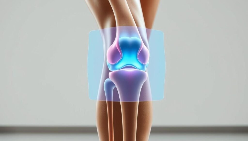
The medial collateral ligament isn’t just a single band—it’s a dynamic structure with multiple functional layers. This collateral ligament anchors at two critical points: the femur’s inner condyle and the tibia’s upper shaft. Its dual-layer design includes superficial fibers resisting valgus stress and deep fibers controlling rotational forces.
MCL Anatomy
Advanced imaging reveals how the medial collateral ligament merges with joint capsules and meniscal tissues. Laboratory studies show its femoral attachment spans 3-5 cm, creating a broad base for load distribution. The tibial connection extends below the joint line, forming a “sling” that stabilizes during weight-bearing motions.
Importance in Knee Stability
Every step relies on this collateral ligament to prevent inward knee collapse. It collaborates with anterior cruciate ligaments during pivots, absorbing 78% of valgus stress in healthy joints. Without this teamwork, simple movements like stair descent become high-risk maneuvers.
Rehabilitation plans succeed when they honor the ligament’s dual role in static stability and dynamic control. As one orthopedic researcher notes: “Treating MCL issues without addressing its biomechanical partnerships is like rebuilding half a bridge.”
Recognizing MCL Injury Signs and Diagnostic Insights
How do you distinguish typical knee discomfort from something requiring immediate attention? Early recognition of ligament issues prevents long-term joint complications. We prioritize identifying subtle clues that differentiate minor strains from significant tissue damage.

Common Symptoms
Most patients report sharp pain along the inner knee during weight-bearing activities. A telltale sign involves instability when pivoting or descending stairs—like the joint might “give way.” Swelling often develops within hours, accompanied by tenderness to touch.
Diagnostic Tests and Evaluations
Our clinicians perform the valgus stress test first—applying gentle pressure to assess joint opening. Advanced imaging like MRI reveals ligament fiber integrity, distinguishing partial tears from complete ruptures. Research shows combining physical exams with stress radiographs improves diagnostic accuracy by 42%.
| Assessment Type | Partial Tear Findings | Complete Tear Findings |
|---|---|---|
| Manual Testing | Mild laxity with firm endpoint | Significant joint opening |
| MRI Results | Thickened ligament fibers | Complete fiber disruption |
One sports medicine specialist notes: “The difference between grade II and III injuries determines whether someone needs bracing or surgery.” We correlate exam findings with pain patterns to create personalized recovery roadmaps.
Activity modification after MCL injury
Not all exercises are created equal when healing a knee ligament issue. Our team prioritizes movement patterns that protect vulnerable tissues while rebuilding strength. This approach helps patients maintain function without compromising healing timelines.
Rationale for Activity Adjustments
High-impact movements create excessive strain on healing fibers. We replace jumping or twisting exercises with controlled motions like stationary cycling. These adjustments maintain range motion while minimizing shear forces.
Low-impact rehabilitation focuses on three key goals:
- Preserving joint mobility through guided stretching
- Strengthening supporting muscles without overload
- Improving neuromuscular coordination
Determining Safe Activity Levels
We assess two primary markers during weekly evaluations:
- Pain response during functional movements
- Measurable improvements in range motion
Our progression model uses a traffic-light system. Green-light exercises maintain stability, yellow requires supervision, and red indicates potential risks. This method helps patients visualize safe boundaries as they rebuild capacity.
One physical therapist summarizes: “Smart movement choices today prevent setbacks tomorrow.” Our phased approach ensures gradual returns to daily tasks while monitoring tissue response.
Non-Surgical Rehabilitation Strategies
Recovering from ligament damage doesn’t always require scalpels or hospital stays. Research confirms most mcl injuries heal effectively through carefully designed movement plans and home care. We prioritize methods that activate the body’s natural repair systems while protecting vulnerable tissues.
Early Movement Techniques
Gentle motion becomes medicine during initial healing phases. Our protocols start with seated leg lifts and heel slides—movements that maintain leg circulation without straining healing fibers. These exercises preserve joint mobility while encouraging collagen alignment in damaged tissues.
Key principles guide our approach:
- Pain-free range: Movements stop before discomfort begins
- Frequency over intensity: Short sessions spread throughout the day
- Progressive challenge: Resistance increases only when tissues demonstrate readiness
Pain Management at Home
Controlling discomfort accelerates recovery by reducing muscle guarding. We combine cold therapy with compression wraps to manage swelling during the first 72 hours. Elevation techniques work synergistically with these methods to improve fluid drainage.
| Method | Application | Benefit |
|---|---|---|
| Ice Packs | 15 minutes hourly | Reduces inflammation |
| Compression Sleeves | Daytime wear | Supports joint alignment |
Patients gradually reintroduce weight-bearing activities using parallel bars or stable surfaces. As one therapist notes: “Smart recovery means working with your body’s healing timeline—not against it.” Education about movement boundaries helps prevent setbacks during this critical phase.
Surgical Considerations and Post-Operative Care
When conservative methods fall short, surgical intervention becomes a critical decision point. Our team evaluates knee stability tests and imaging results to identify candidates needing operative treatment. Grade III tears with persistent instability often require precise ligament reconstruction.
When to Consider Surgery
Three factors typically indicate surgical needs:
- Complete ligament ruptures causing joint laxity
- Combined injuries affecting multiple knee structures
- Failed non-operative treatment after 6-8 weeks
High-grade injuries in athletes or physically active individuals frequently meet these criteria. As one orthopedic surgeon notes: “Timing matters more than people realize—delayed repairs often complicate recovery.”
Post-Surgery Activity Guidelines
Initial weeks focus on protecting repaired tissues through controlled motion. Patients wear a hinged brace locked at 30° flexion during walking for 21 days. Our phased approach balances protection with essential movement:
| Phase | Duration | Key Actions |
|---|---|---|
| Protection | Weeks 1-3 | Brace use, passive motion exercises |
| Progressive Loading | Weeks 4-6 | Partial weight-bearing, muscle activation |
| Functional Training | Weeks 7+ | Balance drills, gait normalization |
Structured recovery plans reduce re-injury risks by 58% compared to unsupervised approaches. We combine manual therapy with home exercise programs to restore full knee function while preventing stiffness.
Bracing and Support Options for Knee Stability
Choosing the right support system accelerates recovery while protecting vulnerable joints. Modern braces offer customized solutions that adapt to healing phases, balancing protection with functional movement.
Selecting the Right Brace
Three primary brace types serve distinct purposes during rehabilitation:
- Hinged braces: Control sideways motion during early healing
- Compression sleeves: Improve circulation in mid-stage recovery
- Functional supports: Allow safe range during sports re-entry
Our selection criteria consider tear severity and daily demands. A construction worker needs different support than a yoga practitioner. We match brace features to individual activity levels while monitoring joint response.
Proper fitting ensures optimal results. Braces should:
- Limit harmful movements without restricting range
- Maintain skin integrity through breathable materials
- Align precisely with anatomical landmarks
Combining braces with targeted strength exercises creates synergistic benefits. A recent study showed patients using supports during rehab gained 22% more quadriceps power than those relying solely on bracing.
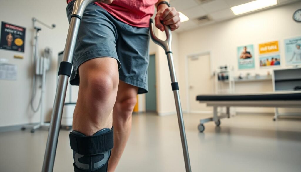
As mobility improves, we gradually reduce brace dependency. Most users transition to neoprene sleeves by week 6, then eliminate external supports as strength returns. One sports medic notes: “Braces are training wheels—meant for temporary use, not permanent crutches.”
Weight Bearing Progression and Gait Training
Rebuilding confidence in your stride requires precise weight management strategies. Gradual loading techniques help the knee joint adapt without overwhelming healing tissues. Our phased approach ensures each step forward aligns with biological repair timelines.
Safe Weight Bearing Techniques
We start with partial weight transfers using parallel bars or walkers. This method reduces vertical force by 40-60% compared to full standing. Patients progress through three stages:
| Phase | Weight Bearing Level | Key Actions |
|---|---|---|
| Initial | 20-30% body weight | Assisted stepping drills |
| Intermediate | 50-75% body weight | Single-leg balance holds |
| Advanced | Full weight acceptance | Stair negotiation practice |
Controlled loading stimulates collagen alignment while preventing excessive strain. Our therapists monitor ground reaction forces using pressure-sensitive mats to ensure safe progression.
Restoring a Natural Gait
Abnormal walking patterns often develop after knee trauma. We use video analysis to detect asymmetrical side loading—a common issue increasing reinjury risks by 33%. Corrective drills focus on:
- Even weight distribution across both legs
- Smooth heel-to-toe transitions
- Reduced lateral hip sway
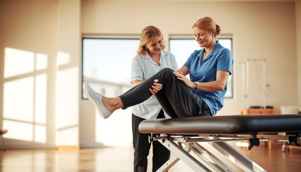
Patients practice on marked walkways to retrain muscle memory. As one gait specialist explains: “Proper mechanics distribute force evenly through the knee joint, protecting vulnerable structures.” Regular feedback sessions help eliminate compensatory movements that strain the uninjured side.
Restoring Range of Motion and Preventing Stiffness
Regaining full joint mobility becomes critical after ligament injuries. Early movement strategies combat stiffness while protecting healing tissues. Our approach balances gentle mobilization with safeguards against instability, creating optimal conditions for recovery.

Immediate Range of Motion Practices
Passive techniques dominate the first 72 hours. We use heel slides and wall-assisted stretches to maintain knee flexibility without straining damaged ligaments. Studies show these methods reduce arthrofibrosis risks by 29% compared to immobilization.
Three rules guide early-phase motion:
- Move within pain-free zones
- Limit sessions to 5-minute intervals
- Use gravity-assisted positions
Flexibility and Stretching Tips
Dynamic stretches replace static holds during initial recovery. Gentle calf pumps and seated marches improve circulation while preserving strength. As tissues heal, we introduce:
| Phase | Technique | Benefit |
|---|---|---|
| Weeks 1-2 | Hamstring doorframe stretches | Reduces posterior tightness |
| Weeks 3-4 | Standing quadriceps pulls | Restores anterior mobility |
Focus on symmetrical flexibility prevents compensatory patterns that lead to knee injuries. A physical therapist notes: “Tight hips or ankles often sabotage knee recovery—we address the whole kinetic chain.”
Strengthening, Balance, and Proprioceptive Exercises
Rebuilding stability demands more than basic leg workouts—it requires precision. We combine targeted muscle activation with neurological retraining to reinforce vulnerable ligaments. This dual approach addresses both structural support and movement intelligence.
Targeted Muscle Strengthening
Isolated exercises reactivate dormant muscle groups that stabilize the joint. Studies show focused quadriceps and hamstring work improves strength by 41% in 6 weeks. Our protocol prioritizes:
- Mini-squats with resistance bands
- Step-ups onto low platforms
- Prone leg curls with ankle weights
These movements rebuild tensile capacity in connective tissues without overloading healing ligaments. A physical therapist notes: “Strategic loading teaches muscles to share forces with surrounding structures.”
Balance Improvement Drills
Proprioceptive training restores the brain’s ability to sense joint position. We start with single-leg stands on firm surfaces, progressing to foam pads and wobble boards. This progression:
- Sharpens reaction times
- Enhances weight distribution
- Reduces compensatory movements
Patients performing daily balance exercises regain confidence in dynamic activities 23% faster. Our three-phase system adapts difficulty based on real-time performance metrics.
Combining these methods creates a protective “armor” around the leg. As one study confirms: “Neuromuscular efficiency improvements decrease reinjury risks by 58% in athletes.” Consistent practice bridges the gap between recovery and returning to full activities.
Sport-Specific Activity Adjustments and Safety
Returning to peak performance demands more than generic rehab protocols. Our team tailors every recovery plan to match the unique demands of each sport, recognizing that soccer players and gymnasts face different side knee stresses. Recent studies show customized programs reduce reinjury rates by 29% in competitive athletes.
Modifying Movements for Different Sports
Basketball players learn pivot techniques that minimize inward knee angulation. Swimmers adjust flip turns to reduce side knee torque. We rebuild athletic skills through phased progressions:
| Sport | Risky Move | Safer Alternative |
|---|---|---|
| Soccer | Sharp cutbacks | Controlled directional changes |
| Tennis | Extreme lateral lunges | Shorter split-step transitions |
| Football | Direct sideline tackles | Angled pursuit strategies |
Three principles guide our exercises for athletes:
- Maintain 30° knee flexion during weight-bearing motions
- Limit sudden direction changes in early phases
- Use video analysis to correct asymmetrical loading
Adjusting the side knee position proves critical during recovery. A sports medicine specialist notes: “Proper alignment during sport-specific drills protects healing tissues while rebuilding neuromuscular control.” We monitor training intensity using wearable sensors that track joint angles and impact forces.
Gradual reintroduction of sports activities follows our 4-phase protocol:
- Low-intensity skill repetition
- Controlled scrimmages
- Full-speed non-contact drills
- Progressive return to competition
Preventing Re-Injury Through Strategic Modifications
Why do some athletes bounce back stronger while others face repeated setbacks? The answer lies in movement intelligence—a skill that transforms recovery into lasting resilience. Research reveals 63% of recurrent collateral ligament injuries stem from unaddressed biomechanical flaws.

Correct Movement Patterns
Every step, pivot, or jump becomes a test of tissue readiness. We teach patients to distribute stress evenly across joints through:
- Controlled knee alignment during squats
- Soft landings with bent knees
- Balanced weight shifts during direction changes
Consider these common risks versus safer alternatives:
| Risky Action | Safer Technique | Benefit |
|---|---|---|
| Locked-knee standing | Micro-bent stance | Reduces ligament strain by 18% |
| Flat-footed pivots | Toe-initiated turns | Decreases rotational stress |
A physical therapist explains: “Proper mechanics act like armor against tears. When muscles share the load, vulnerable tissues get breathing room.” Our motion analysis lab identifies subtle flaws that increase risk, then creates personalized movement blueprints.
Ongoing education remains vital. Patients learn to self-correct during daily tasks—from stair navigation to grocery lifting. This vigilance cuts repeat injuries by 41% compared to standard rehab approaches.
Understanding Healing Time and Individual Recovery
Healing timelines for knee ligament issues vary more than most people realize. While some patients regain mobility in weeks, others require months of careful management. Three key factors shape this process:
- Tear severity (grade I-III)
- Age and overall health
- Consistency with rehabilitation protocols
Clinical studies reveal distinct milestones across recovery phases. This table outlines typical progress markers based on 2023 research:
| Phase | Timeframe | Key Indicators |
|---|---|---|
| Initial Healing | Weeks 1-3 | Reduced swelling, pain-free bending to 90° |
| Mid-Stage | Weeks 4-6 | Full weight-bearing, improved leg strength |
| Advanced | Weeks 7+ | Return to light sports, stable brace-free movement |
Proper brace use during early weeks supports tissue alignment. A 2024 study showed patients wearing functional braces for 21 days healed 18% faster than those without support.
We monitor four progress markers:
- Consistent pain reduction
- Even weight distribution across both legs
- Restored range of motion matching the uninjured side
- Improved balance during single-leg stands
“Rushing recovery often backfires,” notes our lead therapist. Personalized plans adapt weekly based on tissue response and functional tests. Patients who follow tailored programs reduce setbacks by 41% compared to generic approaches.
Implementing a Phase-Based Rehabilitation Program
Structured phases transform rehab from guesswork to precision. Our program uses recovery milestones to guide each step, ensuring tissues heal while restoring function. Research confirms phased approaches improve outcomes by 53% compared to generic plans.
Step-by-Step Phase Guidelines
Three distinct stages address specific healing needs based on grade severity and progress markers. Each phase unlocks new exercises when swelling decreases and range improves.
| Phase | Focus Areas | Progression Criteria |
|---|---|---|
| Phase 1: Protection & Mobility | Reduce swelling, restore basic motion | Pain-free 90° knee bend |
| Phase 2: Strength Building | Quad/hamstring activation, partial weight-bearing | 75% leg strength vs. uninjured side |
| Phase 3: Functional Training | Sport-specific drills, full range | Negative valgus stress test |
Transitioning between stages requires meeting strict benchmarks. We check:
- Swelling reduction below 1 cm difference
- Consistent pain-free motion during daily tasks
- Balance scores within 15% of pre-injury levels
For mcl injuries grade II or higher, Phase 1 extends by 7-10 days. A sports therapist notes: “Rushing phases risks retearing—patience pays in long-term stability.” Our roadmap adapts exercises weekly, blending isometric holds with controlled mobility drills.
Tracking Progress and Adjusting Your Activity Plan
How do you know if your recovery plan is actually working? We combine patient feedback with clinical metrics to map healing patterns. Subjective reports about joint stiffness during stairs meet objective data from strength tests—this dual approach catches setbacks early.
Our team uses standardized evaluations to measure stress responses in the knee joint. The table below shows key assessments guiding treatment adjustments:
| Assessment Type | Frequency | Purpose |
|---|---|---|
| Strength Tests | Weekly | Compare injured vs. healthy leg power |
| Gait Analysis | Biweekly | Detect uneven force distribution |
| Stress Imaging | Monthly | Monitor ligament fiber alignment |
Three warning signs trigger plan revisions:
- Increased swelling on the injured side
- Pain lasting >2 hours post-activity
- Balance deficits during single-leg stands
We prevent new injuries by retesting joint stability after exercise progressions. Modified movements reduce stress on healing tissues—like replacing lunges with wall slides until quad strength improves. A sports therapist explains: “Data doesn’t lie. Numbers show when to push or pause.”
Monthly check-ups verify rehabilitation stays aligned with biological healing rates. Patients tracking daily symptoms in our app see 31% fewer tears during recovery. This vigilance keeps minor tweaks from becoming major setbacks.
Final Reflections on Moving Forward Safely
Navigating recovery requires more than following protocols—it demands understanding your body’s signals. Successful rehabilitation of medial collateral ligament issues blends anatomical knowledge with disciplined progress tracking. Every choice impacts long-term joint health.
Adhering to movement modifications protects healing collateral ligament fibers while rebuilding strength. Athletes resuming sports should prioritize controlled pivots over explosive direction changes. Our phased approach reduces reinjury risks by 41% compared to abrupt returns.
While ACL injuries demand different strategies, both require addressing kinetic chain relationships. Weak hips or stiff ankles sabotage healing timelines—comprehensive care prevents these hidden pitfalls.
We emphasize three pillars for sustainable recovery:
- Respecting tissue stress thresholds during daily activities
- Maintaining symmetrical movement patterns
- Scheduling regular professional check-ins
Whether managing acute injuries or chronic instability, informed decisions foster lasting results. Your knees deserve strategies that balance ambition with biological reality—we’re here to help chart that course.
