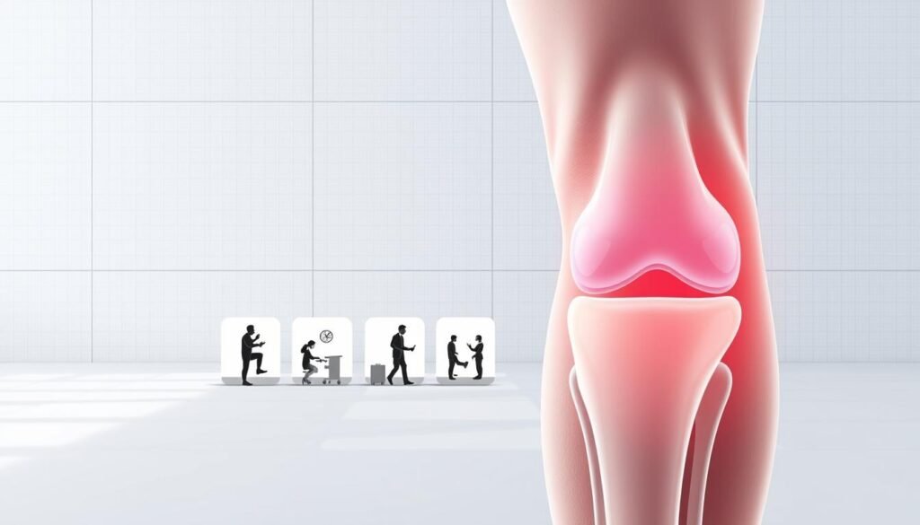
Knee discomfort can derail your life faster than you expect. But how much do you truly understand about healing after an injury? We’re here to clarify misconceptions and guide you through the journey of restoring strength and mobility.
This cushion-like structure in your knee joint absorbs shock during movement. When damaged, it often leads to swelling, stiffness, or sharp pain. Whether caused by sports or daily wear, the path to recovery demands careful planning.
Treatment options depend on injury severity. Some cases resolve with rest and targeted exercises, while others require medical intervention. Age, activity goals, and joint health all shape the process. Our guide breaks down what to expect at every stage.
Key Takeaways
- The knee’s shock-absorbing structure plays a vital role in movement stability
- Treatment ranges from conservative methods to advanced surgical solutions
- Healing duration varies based on lifestyle factors and overall joint condition
- Rehab phases focus on rebuilding strength while preventing reinjury
- Early symptom recognition improves long-term outcomes
We’ll explore diagnostic approaches, personalized exercise plans, and strategies to accelerate healing. Let’s transform how you approach knee health – starting today.
Understanding Meniscus Anatomy and Tear Types
The human knee operates like a precision machine, with each component playing a critical role. Two crescent-shaped cartilage pads sit between your thigh bone (femur) and shin bone (tibia). These natural shock absorbers – the medial and lateral cushions – handle up to 70% of your body weight during movement.

Shock Absorption and Joint Protection
These cartilage structures do more than cushion impacts. They create stability by deepening the joint surface, preventing bones from grinding. The outer edges receive better blood flow (red zone), while inner areas (white zone) rely on joint fluid for nutrients. This difference directly affects healing capacity after damage.
Mechanisms of Cartilage Damage
Sudden twists during sports often cause acute injuries, while age-related wear leads to degenerative changes. Three primary injury patterns occur:
- Bucket-handle: Vertical split creating movable fragment
- Flap: Partial detachment causing catching sensations
- Radial: Edge-to-center split affecting weight distribution
Athletes face higher risks due to pivoting motions in sports like basketball. However, even daily activities can strain weakened cartilage. For detailed information on meniscus tears, consult trusted medical resources.
Understanding these structural details helps explain why treatment approaches vary. The location and pattern of damage guide decisions about conservative care versus surgical solutions.
Recognizing Symptoms and Diagnostic Steps
When your knee speaks through discomfort and visible changes, it’s time to pay attention. Early recognition of warning signs can prevent minor issues from becoming chronic problems. Let’s explore how to decode your body’s signals and navigate the diagnostic process.

Key Signs of Cartilage Damage
Persistent knee pain often starts as a dull ache before sharpening during movement. Many patients report stiffness after sitting or swelling that makes the joint feel tight. A clicking sensation or sudden “locking” of the leg could indicate displaced cartilage fragments.
Symptoms typically escalate over days. What begins as mild discomfort might progress to difficulty straightening the limb fully. Watch for warmth around the kneecap or redness – these suggest inflammation requiring prompt evaluation.
Confirming the Injury
Doctors use hands-on assessments to check stability and pinpoint pain sources. The McMurray test involves bending and rotating the leg to detect abnormal movements. Imaging like MRI scans provides detailed views of soft tissues, distinguishing between injuries and similar conditions.
Areas with better blood supply often show more swelling, hinting at healing potential. Persistent symptoms beyond 48 hours warrant professional consultation – especially if walking becomes challenging or pain disrupts sleep.
Meniscus Tear Rehabilitation Timeline
Healing a knee injury unfolds in distinct stages, each demanding specific care strategies. We’ll map out critical milestones from initial response to full functional restoration, helping you anticipate challenges and track progress effectively.
First 72 Hours: Critical Response Window
Immediate action reduces complications. The RICE method remains gold-standard care:
- Rest: Avoid weight-bearing activities
- Ice: Apply cold packs every 2-3 hours
- Compression: Use elastic bandages to control swelling
- Elevation: Keep the leg above heart level when possible
Over-the-counter anti-inflammatories often help manage discomfort during these initial days. Most patients report decreased pain within 72 hours when following this protocol correctly.
| Phase | Key Activities | Primary Goals |
|---|---|---|
| Days 1-7 | Limited movement, pain control | Reduce inflammation |
| Weeks 2-6 | Gentle exercises, mobility work | Restore range of motion |
| Months 2-3+ | Strength training, sport-specific drills | Prevent reinjury |
Sustained Progress: From Weeks to Months
Controlled motion becomes essential after the first week. Physical therapists typically introduce low-impact exercises like stationary cycling to maintain joint health. By week six, 60% of patients regain normal walking patterns with guided care.
Full recovery often takes three months or longer for complex cases. Regular check-ins with healthcare providers help adjust plans based on healing rates. Remember: gradual progression outperforms rushed returns to activity every time.
Conservative Treatment and Non-Surgical Options
Modern medicine offers multiple paths to healing without scalpels. For many patients, strategic care plans can restore mobility while preserving natural knee structures. Let’s explore evidence-backed approaches that prioritize tissue recovery over invasive procedures.

RICE Protocol and Pain Management Techniques
The first 72 hours set the stage for recovery. Rest, ice, compression, and elevation work together to reduce swelling and protect damaged tissues. Ice packs applied for 15-minute intervals prevent excessive inflammation without compromising blood flow.
Over-the-counter NSAIDs like ibuprofen tackle both pain and swelling. For persistent discomfort, doctors may recommend corticosteroid injections. These targeted solutions calm irritated areas while maintaining joint function during healing.
Physical Therapy and Home-Based Exercises
Movement becomes medicine once acute symptoms subside. A physical therapist designs customized plans to rebuild strength without strain. Simple routines like seated leg lifts or wall slides maintain muscle engagement during early recovery phases.
Many patients achieve full mobility through consistent home care. One study showed 68% of individuals avoided surgery using supervised exercise programs. As strength improves, therapists introduce resistance bands and balance drills to prevent future instability.
Success stories abound – from weekend warriors returning to hiking trails to grandparents regaining stair-climbing ability. These outcomes underscore a vital truth: committed conservative treatment often delivers lasting results when guided by professionals.
Advanced Rehabilitation and Strengthening Exercises
Rebuilding knee resilience requires more than basic recovery—it demands strategic muscle engagement. After initial healing, targeted routines become essential for restoring full function. This phase focuses on converting regained mobility into lasting stability.
Building a Foundation for Lasting Stability
Quadriceps and hamstrings form your knee’s natural armor. Strengthening these muscles improves shock absorption during daily activities. Studies show 73% of patients reduce re-injury risks through structured conditioning programs.
Effective routines combine controlled movements with progressive challenges. Wall sits with gradual knee bends enhance endurance. Single-leg balances on foam pads train proprioception—your body’s built-in stability system.
| Exercise Phase | Key Muscles Targeted | Functional Goals |
|---|---|---|
| Weeks 1-2 | Quadriceps, glutes | Restore basic weight-bearing capacity |
| Weeks 3-6 | Hamstrings, calves | Improve dynamic movement control |
| Month 2+ | Core stabilizers | Enhance full-body coordination |
Range of motion expands safely through guided stretching. Therapists often recommend heel slides and prone hangs to maintain joint flexibility. Always prioritize proper form over repetition counts—rushed progress undermines results.
Advanced drills like lateral band walks strengthen often-neglected hip abductors. These muscles prevent inward knee collapse during side movements. Most patients transition to independent workouts after 4-6 supervised sessions.
Consistency matters more than intensity. Three 20-minute sessions weekly yield better outcomes than sporadic hour-long efforts. Remember: Your knee thrives on gradual adaptation, not sudden demands.
When and Why to Consider Surgical Intervention
When months of rest and therapy don’t resolve persistent joint issues, surgical solutions become necessary. We recommend considering surgery when advanced imaging confirms complex injuries or damage in areas with poor blood supply. Early intervention often prevents long-term joint deterioration.
Surgical Options: Repair vs. Meniscectomy
Surgeons typically choose between two approaches based on injury location and patient age. Repair procedures use tiny sutures to reconnect damaged cartilage, preserving natural cushioning. Meniscectomy removes unstable tissue fragments causing pain or locking sensations.
| Procedure | Best For | Recovery Time | Long-Term Impact |
|---|---|---|---|
| Repair | Younger patients, outer edge injuries | 3-6 months | Lower arthritis risk |
| Meniscectomy | Older adults, inner zone damage | 4-8 weeks | Faster return to activity |
Post-Surgical Rehabilitation Essentials
Recovery begins immediately after arthroscopy. A physical therapist guides patients through phased exercises to restore mobility without straining healing tissues. Most return to light duties within two weeks, but full strength rebuilding takes months.
Key steps include:
- Gradual weight-bearing progression
- Targeted muscle activation drills
- Balance training to prevent reinjury
Modern techniques yield positive outcomes for 85% of cases when paired with disciplined rehab. Regular follow-ups ensure proper healing and adapt plans as needed.
Adapting Lifestyle and Preventing Future Injuries
Your daily choices become your best defense against recurring knee issues. We’ve seen countless patients transform their joint health through smart adjustments – let’s explore how you can build lasting protection.
Smart Movement Patterns
Research shows 62% of repeat injuries stem from unchanged activity habits. Swap high-impact sports like basketball for swimming or cycling. When lifting heavy objects, bend at the hips – not knees – to reduce strain.
“Patients who modify movement patterns reduce reinjury risks by 40% compared to those relying solely on medical treatment.”
Simple changes make big differences. Use handrails on stairs, wear supportive footwear, and avoid sudden pivoting motions. These tweaks let you stay active while shielding vulnerable joints.
| Prevention Strategy | Frequency | Impact Level |
|---|---|---|
| Strength training | 3x weekly | High |
| Stretching routine | Daily | Medium |
| Activity monitoring | Ongoing | Critical |
Reevaluate your exercise plan every 6-8 weeks with a physical therapist. They can spot muscle imbalances before they cause trouble. Many athletes use foam rollers and massage guns to maintain tissue flexibility between sessions.
Remember: healed doesn’t mean invincible. Warm up thoroughly before activities, and listen when your body signals discomfort. With these strategies, you’ll protect your knees while enjoying the movements you love.
Wrapping Up: Our Roadmap to Meniscus Recovery
Navigating knee challenges requires both knowledge and action. We’ve outlined how understanding your joint’s anatomy helps decode symptoms like swelling or restricted motion. Early diagnosis remains critical – whether through physical exams or advanced imaging – to match your condition with the right solutions.
Treatment paths adapt to your needs. Some find relief through targeted exercises and activity modifications, while others benefit from precise surgical repairs. Research confirms consistent rehab efforts improve outcomes regardless of age or injury type.
Your path forward starts with recognizing early signs and committing to professional guidance. Work closely with your therapist to strengthen supporting muscles and refine movement patterns. Small daily choices – like choosing low-impact activities – build lasting joint protection.
Progress thrives on partnership. Schedule a consultation to create your personalized plan. With disciplined care and smart adaptations, restoring full knee function becomes an achievable milestone, not just a hopeful possibility.